Can dwarfism be detected on ultrasound
By David Edwards
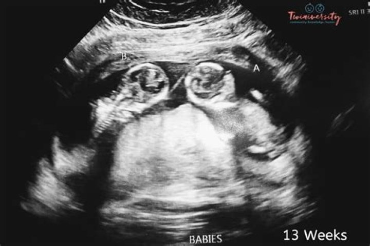
Doctors are able to diagnose most cases of achondroplasia even before birth by doing an ultrasound in the later stages of pregnancy. The ultrasound can show if a baby’s arms and legs are shorter than average and if the baby’s head is larger.
Can you tell if your baby has dwarfism in the womb?
Imaging. Doctors may spot signs of achondroplasia, such as shorter limbs, or other causes of dwarfism on ultrasounds of a fetus during pregnancy. X-rays of babies or children may show that their arms or legs are not growing at a normal rate, or that their skeleton shows signs of dysplasia.
When does achondroplasia appear?
Achondroplasia appears to affect males and females in equal numbers. This disorder begins in the developing fetus and is one of the most common forms of skeletal dysplasia that causes dwarfism. The estimated frequency of achondroplasia has ranged from about one in 15,000 to one in 35,000 births.
What are signs of dwarfism in ultrasound?
- a larger head with a prominent forehead.
- a flattened bridge of the nose.
- shortened hands and fingers.
- a sway of the lower back.
- bowed legs.
When does achondroplasia appear in ultrasound?
Discussion. Nowadays, achondroplasia is suspected only after the third trimester due to the late appearance of this disease [2]. These fetuses almost always have a long bone below the third percentile for gestational age but normal size of head and abdominal circumference [2], [8], [9], [10].
Are there prenatal tests for achondroplasia?
Achondroplasia is generally detected by abnormal prenatal ultrasound findings in the third trimester of pregnancy. It is then confirmed by molecular genetic testing of fetal genomic DNA obtained by percutaneous umbilical blood sampling (PUBS).
How is achondroplasia diagnosed ultrasound?
Achondroplasia can be diagnosed before birth by fetal ultrasound. This test uses sound waves and a computer to create images of the baby growing in the womb. DNA testing can also be done before birth to confirm fetal ultrasound results. The condition can also be diagnosed after birth with a physical exam.
Can NIPT detect achondroplasia?
Flowchart summarizing non-invasive prenatal testing (NIPT) results for fetal achondroplasia in high-risk pregnancies and low-risk controls.Can two normal parents have a child with achondroplasia?
When both parents have achondroplasia, the chance for them, together, to have a child with normal stature is 25 percent. Their chance of having a child with achondroplasia is 50 percent.
What are Trident hands?Definition. A hand in which the fingers are of nearly equal length and deflected at the first interphalangeal joint, so as to give a forklike shape consisting of separation of the first and second as well as the third and fourth digits. [ from HPO]
Article first time published onAre there any cures for achondroplasia?
Currently there are no treatments able to reverse achondroplasia, which is caused by mutations in a gene — called FGFR3 — that result in the excess production of proteins that slow bone growth, nor are there ways to treat the genetic culprit itself.
How can achondroplasia be diagnosed?
Achondroplasia Diagnosis Achondroplasia can be diagnosed before birth by fetal ultrasound or after birth by complete medical history and physical examination. DNA testing is now available before birth to confirm fetal ultrasound findings for parents who are at increased risk of having a child with achondroplasia.
Does NIPT test include ultrasound?
Since NIPT is a blood test for a specific disorder, it does not provide a comprehensive evaluation of your baby. A comprehensive test should include not only blood work but an Ultrasound to assess your baby’s anatomy.
Does NIPT include ultrasound?
This combines results from a blood test, the mother’s age and an ultrasound scan (which measures the thickness of fluid behind the baby’s neck, called the nuchal translucency) to show whether the baby is at increased risk of Down syndrome.
Do you have an ultrasound with NIPT?
We recommend that prior to having NIPT, the woman has an ultrasound to confirm the gestation of the pregnancy, the number of fetuses and that the pregnancy is viable. Please note that NIPT is not a test of fetal viability as a demised fetus will continue to release DNA into the mother’s circulation.
What is Rhizomelic?
Abstract. The term rhizomelic pertains to the proximal portions of the limbs including shoulder and arm in the upper extremity and hip and thigh in the lower extremity.
What causes achondroplasia?
Mutations in the FGFR3 gene cause achondroplasia. The FGFR3 gene provides instructions for making a protein that is involved in the development and maintenance of bone and brain tissue. Two specific mutations in the FGFR3 gene are responsible for almost all cases of achondroplasia.
What does Hypochondroplasia mean?
Hypochondroplasia is a form of short-limbed dwarfism. This condition affects the conversion of cartilage into bone (a process called ossification), particularly in the long bones of the arms and legs.
Does HGH Help achondroplasia?
Growth hormone is used to increase the height of patients with achondroplasia (see Medical Care). However, no long-term studies exist to justify prolonged treatment for short stature.
What is the life expectancy for a person with achondroplasia?
AchondroplasiaTreatmentSupport groups, growth hormone therapy, treatment of complicationsPrognosis10-year shorter life expectancyFrequency1 in 27,500 people
How accurate is NIPT for gender?
The chances of a sex determination via NIPT being wrong is around 1 percent when the test is conducted after week 10 of your pregnancy or later, Schaffir says.
What is a first trimester screening ultrasound?
What is first trimester screening? First trimester screening combines fetal ultrasound and blood tests for the mother. It’s done during the first trimester of pregnancy, during weeks 1 to 12 or 13. It can help find out the risk of the baby having certain birth defects.
How many ultrasounds do you have during your pregnancy?
Most healthy women receive two ultrasound scans during pregnancy. “The first is, ideally, in the first trimester to confirm the due date, and the second is at 18-22 weeks to confirm normal anatomy and the sex of the baby,” explains Mendiola.
What makes you high risk for Down's syndrome baby?
One factor that increases the risk for having a baby with Down syndrome is the mother’s age. Women who are 35 years or older when they become pregnant are more likely to have a pregnancy affected by Down syndrome than women who become pregnant at a younger age.
Is NIPT or ultrasound more accurate?
Other research has shown that NIPTs are more accurate than those same standard screenings in predicting the risk of Down syndrome (NIPTs are 99 percent accurate) and Edwards syndrome.
What's the difference between NIPT and NT?
The NIPT by GenePlanet test is much more accurate than the nuchal translucency scan. Its detection rate for the three most common trisomies present at birth is higher than 99%. for the most common trisomies, 0.14% overall.