How do upper motor neurons work
By David Edwards
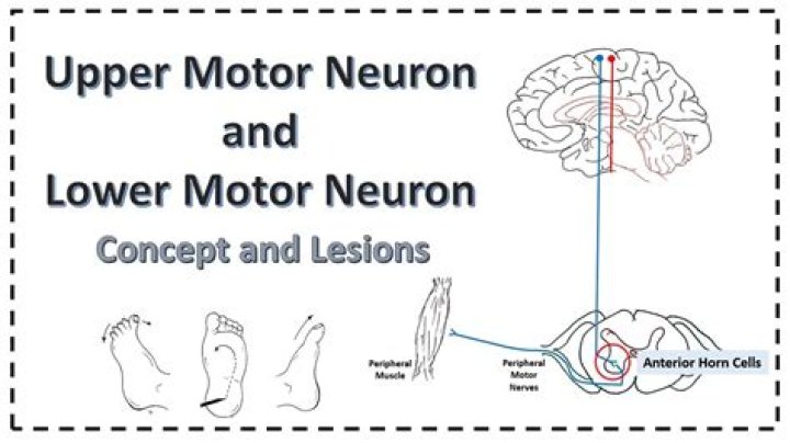
Upper motor neurons are located in your brain and spinal cord. They send signals to lower motor neurons. Lower motor neurons are in your brain stem and spinal cord. When they get a signal from the upper motor neurons, they send another signal to your muscles to make them contract.
How do upper and lower motor neurons work?
The upper and lower motor neurons form a two-neuron circuit. The upper motor neurons originate in the cerebral cortex and travel down to the brain stem or spinal cord, while the lower motor neurons begin in the spinal cord and go on to innervate muscles and glands throughout the body.
What do upper motor neurons release?
Upper motor neuronFMA84631Anatomical terminology
What is the difference between upper and lower motor neuron lesions?
An upper motor neuron lesion is a lesion of the neural pathway above the anterior horn of the spinal cord or motor nuclei of the cranial nerves. A Lower motor neuron lesion is a lesion which affects nerve fibers traveling from the anterior horn of the spinal cord to the associated muscle(s).What is the target of an upper motor neuron?
The target of the upper motor neuron is the dendrites of the lower motor neuron in the gray matter of the spinal cord. (8) The axon of the lower motor neuron emerges from the spinal cord in a nerve and connects to a muscle through a neuromuscular junction to cause contraction of the target muscle.
What is one primary difference between an upper motor neuron and a lower motor neuron?
Upper Motor NeuronLower Motor NeuronIt is entirely located in the central nervous systemIt is either located in the spinal cord gray matter or within the cranial nerve nuclei in the brain stem.
Why is UMN spastic?
How does UMN lesion cause spasticity and associated phenomena? The major problem is a loss of control of the spinal reflexes. Spinal reflex activity is normally tightly regulated and if inhibitory control is lost, the balance is tipped in favor of excitation, resulting in hyperexcitability of the spinal reflexes.
Where do upper motor neuron lesions occur?
Upper motor neuron lesions occur in the brain or the spinal cord as the result of stroke, multiple sclerosis, traumatic brain injury, cerebral palsy, atypical parkinsonisms, multiple system atrophy, and amyotrophic lateral sclerosis.What are upper neurons?
Upper motor neurons are located in your brain and spinal cord. They send signals to lower motor neurons. Lower motor neurons are in your brain stem and spinal cord. When they get a signal from the upper motor neurons, they send another signal to your muscles to make them contract.
Why does UMN lesion increase tone?Muscle tone is increased in upper motor neuron lesions, for example in cerebral cortical damage that occurs in cerebrovascular accident. This is thought to be due to loss of cortical control of motor neurons, which increase their activity.
Article first time published onWhat is UMN lesion?
UMN lesions are designated as any damage to the motor neurons that reside above nuclei of cranial nerves or the anterior horn cells of the spinal cord. Damage to UMN’s leads to a characteristic set of clinical symptoms known as the upper motor neuron syndrome.
Why is there Hyperreflexia in UMN lesions?
Hyperreflexia. Because of the loss of inhibitory modulation from descending pathways, the myotatic (stretch) reflex is exaggerated in upper motor neuron disorders. The stretch reflex is a major clinical diagnostic test of whether a motor disorder is caused by damage to upper or lower motor neurons.
Why are upper motor neuron lesions characterized by spastic paralysis?
Why are upper motor neuron lesions characterized by “spastic paralysis”? A. Paralysis means that voluntary muscle control is not possible because of the interruption of descending motor input. Spasticity refers to what could be called “hypercontractility” of the muscles in the absence of the descending input.
Are upper motor neurons efferent?
The axons of lower motor neurons are efferent nerve fibers that carry signals from the spinal cord to the effectors. Upper motor neurons are corticospinal interneurons that arise from the motor cortex of the brain and descend into the spinal cord where they activate lower motor neurons through synapses.
What contains upper motor neuron cell?
The cell bodies of these neurons are located within the ventral horns of the spinal cord and within brainstem motor nuclei. Upper motor neurons, as defined clinically, are cortical neurons that innervate lower motor neurons (either directly or via local interneurons).
What might a reflex look like in someone with an upper motor neuron lesion?
Patients can be seen to have abnormally brisk reflexes which are due to decreased modulation by descending inhibitory pathways. Radiation of reflexes is a regular observation with the hyperreflexia of UMN lesions.
What is a clonus?
Clonus is involuntary and rhythmic muscle contractions caused by a permanent lesion in descending motor neurons. Clonus may be found at the ankle, patella, triceps surae, wrist, jaw, biceps brachii.
What causes clonus?
Clonus results due to an increased motor neuron excitation (decreased action potential threshold) and is common in muscles with long conduction delays, such as the long reflex tracts found in distal muscle groups. Clonus is commonly seen in the ankle but may exist in other distal structures as well.
Is ALS UMN or LMN?
Typical, or “classical,” ALS is associated with simultaneous upper motor neuron (UMN) and lower motor neuron (LMN) involvement at disease onset, whereas atypical forms, such as primary lateral sclerosis and progressive muscular atrophy, have early and predominant involvement in the UMN and LMN, respectively.
What causes Hyperreflexia and clonus?
Common causes of hyperreflexia include focal brain lesions (typically causing unilateral hyperreflexia), cervical myelopathy, and motor neuron disease (amyotrophic lateral sclerosis, ALS). The latter is characterized by a combination of upper and lower motor neuron findings.
Are upper motor neurons inhibitory?
Upper motor neurons may stimulate or inhibit motor actions. For instance, the UMN is necessary for voluntary motor action and at the same time exerts inhibitory action on other functions, such as spinal cord reflexes.
Why is there hypotonia in UMN lesions?
This initial period of “hypotonia” after upper motor neuron injury is called spinal shock, and reflects the decreased activity of spinal circuits suddenly deprived of input from the motor cortex and brainstem.
Is clonus an upper motor neuron lesion?
Clonus is a rhythmic oscillating stretch reflex that is related to upper motor neuron lesions. Therefore, clonus is generally accompanied by hyperreflexia.
What is the role of acetylcholine at the neuromuscular junction?
Acetylcholine is a small molecule that acts as a chemical messenger to propagate nerve impulses across the neuromuscular junction between a nerve and a muscle. … And it is this sodium that regenerates the nerve impulse in the muscle fibre and makes it contract.
What is Hyperreflexia and clonus?
Clonus is the highest degree of hyperreflexia. The most important neuromuscular disease associated with hyperreflexia is ALS due to degeneration of the cortical motor neurons. Diagnostic difficulty occurs when hyperreflexia and spasticity are the only findings.
What role does dopamine play in motor control?
Dopamine is a specialised messenger molecule that alters the way other neurons processes information. In the normal brain, dopamine controls information processing in neurons that affect movement, attention and motivation.
What is the target of an upper motor neuron quizlet?
What is the target of an upper motor neuron? sodium and potassium ions inside the cell.
Which is a difference between summation and tetanus?
Summation and Tetanus Contractions: Repeated twitch contractions, where the previous twitch has not relaxed completely are called a summation. If the frequency of these contractions increases to the point where maximum tension is generated and no relaxation is observed then the contraction is termed a tetanus.
What are upper and lower motor neuron symptoms?
Upper motor neuron disease causes stiffness, which is called “spasticity”. Lower motor neuron disease causes weakness, loss of muscle (“atrophy”) and muscle twitching (“fasciculations”).
What are examples of interneurons?
In human brain, there are about 100 billion interneurons. Example is the Golgi cell found in the cerebellum. The interneurons receive impulses from the sensory neurons. They interpret the information received from other neurons and relay impulses to motor neurons for an appropriate response.
Can upper motor neurons regenerate?
Motor neurons, which have processes that reside in both the CNS and the PNS, do regenerate, however. In the absence of intervention, motor neurons are one of the only CNS neurons to regenerate following axotomy.