What are lamina in spinal cord
By Christopher Green
The lamina is the flattened or arched part of the vertebral arch, forming the roof of the spinal canal; the posterior part of the spinal ring that covers the spinal cord or nerves.
How many lamina is present in spinal cord?
structure of spinal cord The gray matter of the spinal cord is composed of nine distinct cellular layers, or laminae, traditionally indicated by Roman numerals.
What is brain lamina?
The lamina terminalis is a thin sheet of gray matter and pia mater that attaches to the upper surface of the chiasm and stretches upward to fill the interval between the optic chiasm and the rostrum of the corpus callosum.
What is lamina II?
Lamina II is involved in the reception of all sensory-related stimuli (both noxious and non-noxious), and specifically moderates the sensation of pain. This lamina maintains direct communication with Laminae III and IV and corresponds to the spinal cord nucleus known as the substantia gelatinosa.What is lamina Rexed?
Rexed’s laminae is an architectural classification of the structure of the spinal cord, based on the cytological features of the neurons in different regions of the gray substance described by the Swedish Anatomist B. Rexed.
What is a Dermatome?
A dermatome is an area of skin in which sensory nerves derive from a single spinal nerve root (see the following image). Dermatomes of the head, face, and neck. … Sensory information from a specific dermatome is transmitted by the sensory nerve fibers to the spinal nerve of a specific segment of the spinal cord.
Where is lamina in leaf?
The flat and expanded portion of the leaf in its entirety is known as the lamina. In short, the blade of the leaf or the leaf blade is called the lamina of a leaf.
What is posterior root ganglion?
A dorsal root ganglion (or spinal ganglion; also known as a posterior root ganglion) is a cluster of neurons (a ganglion) in a dorsal root of a spinal nerve. The cell bodies of sensory neurons known as first-order neurons are located in the dorsal root ganglia.What is the anatomy of the spinal cord?
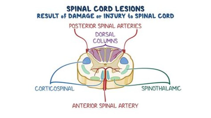
The spinal cord is a cylindrical structure of nervous tissue composed of white and gray matter, is uniformly organized and is divided into four regions: cervical (C), thoracic (T), lumbar (L) and sacral (S), (Figure 3.1), each of which is comprised of several segments.
What is the tract of Lissauer?Anatomical terms of neuroanatomy The posterolateral tract (fasciculus of Lissauer, Lissauer’s tract, tract of Lissauer, dorsolateral fasciculus, dorsolateral tract, zone of Lissauer) is a small strand situated in relation to the tip of the posterior column close to the entrance of the posterior nerve roots.
Article first time published onWhat is lamina in biology?
Lamina is a general anatomical term meaning “plate” or “layer”. It is used in both gross anatomy and microscopic anatomy to describe structures. … The vertebral laminae: plates of bone that form the posterior walls of each vertebra, enclosing the spinal cord. The laminae of the thalamus: the layers of thalamus tissue.
What surrounds the central canal of the spinal cord?
The spinal cord (and brain) are protected by three layers of tissue or membranes called meninges, that surround the canal. The dura mater is the outermost layer, and it forms a tough protective coating. Between the dura mater and the surrounding bone of the vertebrae is a space called the epidural space.
Where is the interventricular foramen?
The interventricular foramen is located between the thalamus and anterior pillar of the fornix, at the anterior margin of the body. The 2 interventricular foramens (or foramina of Monro) connect the lateral ventricles with the third ventricle.
What is white and GREY matter in the spinal cord?
The central nervous system, which consists of the brain and spinal cord, contains two types of tissue. … Gray matter contains neural cells, dendrites, and axon terminals, while white matter consists of axons and myelin, and plays a key role in nerve cells’ ability to connect to one another.
What is lamina Marginalis?
The marginal nucleus of spinal cord, or posteromarginal nucleus, Rexed lamina I, is located at the most dorsal aspect of the dorsal horn of the spinal cord. The neurons located here receive input primarily from Lissauer’s tract and relay information related to pain and temperature sensation.
What is the function of ventral horn?
The ventral horns contains the cell bodies of motor neurons that send axons via the ventral roots of the spinal nerves to terminate on striated muscles.
What are the function of Lamina?
The lamina is the expanded portion or blade of a leaf and it is an above-ground organ specialized for photosynthesis.
What is called lamina in leaf?
Lamina of the leaf (Blade) [ Botany] The lamina is the expanded portion or blade of a leaf and it is an above-ground organ specialized for photosynthesis.
What is the main function of leaf lamina?
Photosynthesis is the main function of leaf lamina.
What are examples of Dermatomes?
Nerve RootDermatomesCervicalL3Back, upper buttock, anterior thigh and knee, medial lower legL4Medial buttock, latera thigh, medial leg, dorsum of foot, big toeL5Buttock, posterior and lateral thigh, lateral aspect of leg, dorsum of foot, medial half of sole, first, second, and third toes
What are dermatome levels?
Rationale. The area of skin that is supplied by a single spinal nerve is known as a dermatome. … The area of sensory block should be assessed using cold sensation (eg ice) to establish which dermatome levels are covered. Both left and right sides need to be assessed.
What is the clinical significance of the dermatome?
Dermatomes are useful to help localize neurologic levels, particularly in radiculopathy. Effacement or encroachment of a spinal nerve may or may not exhibit symptoms in the dermatomic area covered by the compressed nerve roots in addition to weakness, or deep tendon reflex loss.
What are the 5 sections of the spine?
The spine is composed of 33 bones, called vertebrae, divided into five sections: the cervical, thoracic, and lumbar spine sections, and the sacrum and coccyx bones. The cervical section of the spine is made up of the top seven vertebrae in the spine, C1 to C7, and is connected to the base of the skull.
What does S1 and S2 nerve control?
The sacral spine consists of five segments, S1 – S5, that together affect nerve communication to the lower portion of the body. … S1 affects the hips and groin area. S2 affects the back of the thighs. S3 affects the medial buttock area.
Why does L2 end spinal cord?
It is these spinal nerve roots that compose the cauda equina beyond L1/L2. The fact that the spinal cord ends at L1/L2 is very useful in clinical practice in that it allows for spinal taps to be performed to sample CSF without the risk of puncturing the spinal cord.
Where is the spinal ganglion located?
Introduction. The dorsal root ganglia (or spinal ganglia) are described as nodule-like structures found on the posterior roots of each spinal nerve, which contain the soma (or cell bodies) of the afferent sensory nerves carrying sensory signals back to the central nervous system (Figure 33.1) (Standring, 2008).
What happens when the dorsal root ganglia is damaged?
Damage to the dorsal root ganglion cells leads to simultaneous degeneration of short (non- length dependent) as well as long (length dependent) axons and it is this feature that is the key to understanding the clinical pre- sentation.
What is ventral root ganglion?
Each spinal cord segment has two ventral roots that connect by a white ramus to a spinal sympathetic ganglion. These ganglia communicate with each other up and down the spinal cord, forming two sympathetic chains, one on each side of the vertebral column.
What does zone of Lissauer do?
The fibers in the dorsolateral fasciculus (tract of Lissauer or lateral spinothalamic tract), which are concerned with pain, temperature, and light touch, synapse in the substantia gelatinosa, decussate, and then ascend in the lateral column of the spinal cord and brainstem to the thalamus.
Where does the spinothalamic tract terminate?
The spinothalamic tract terminates mainly in the ventroposterolateral nucleus, ventroposteromedial nucleus, the intralaminar nuclei, mainly the central lateral nucleus, and the posterior complex.
What is a lamina answer?
Answer: a thin layer, plate, or scale of sedimentary rock, organic tissue, or other material. Explanation: Lamina is a general anatomical term meaning “plate” or “layer”. … Some examples include: The laminae of the thyroid cartilage: two leaflike plates of cartilage that make up the walls of the structure.