What are the components of ECG
By James Craig
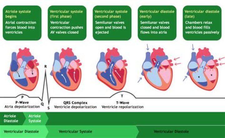
There are three main components to an ECG: the P wave, which represents the depolarization of the atria; the QRS complex, which represents the depolarization of the ventricles; and the T wave, which represents the repolarization of the ventricles.
What are the 5 components of an ECG?
- P wave. The P wave is a small deflection wave that represents atrial depolarization.
- PR interval. …
- QRS wave complex. …
- ST segment. …
- T wave. …
- Wave direction and size. …
- Interpreting the ECG. …
- Rate.
What is aVR aVL and aVF in ECG?
aVR means augmented Vector Right; the positive electrode is on the right shoulder. aVL means augmented Vector Left; the positive electrode is on the left shoulder. aVF means augmented Vector Foot; the positive electrode is on the foot.
What is shown on an ECG?
An ECG (electrocardiogram) records the electrical activity of your heart at rest. It provides information about your heart rate and rhythm, and shows if there is enlargement of the heart due to high blood pressure (hypertension) or evidence of a previous heart attack (myocardial infarction).Which of the following components of an ECG represents atrial depolarization?
The P wave represents the depolarization of the left and right atrium and also corresponds to atrial contraction.
What is abnormal ECG?
An abnormal ECG can mean many things. Sometimes an ECG abnormality is a normal variation of a heart’s rhythm, which does not affect your health. Other times, an abnormal ECG can signal a medical emergency, such as a myocardial infarction /heart attack or a dangerous arrhythmia.
What is a normal ECG reading?
Normal intervals Normal range 120 – 200 ms (3 – 5 small squares on ECG paper). QRS duration (measured from first deflection of QRS complex to end of QRS complex at isoelectric line). Normal range up to 120 ms (3 small squares on ECG paper).
What does V1 V2 V3 mean in ECG?
The areas represented on the ECG are summarized below: V1, V2 = RV. V3, V4 = septum. V5, V6 = L side of the heart. Lead I = L side of the heart.What are the 2 main things an ECG can tell you about a patient's heart?
An ECG records these impulses to show how fast the heart is beating, the rhythm of the heart beats (steady or irregular), and the strength and timing of the electrical impulses as they move through the different parts of the heart. Changes in an ECG can be a sign of many heart-related conditions.
What does AVL measure?LabelMeaning of labelPosition of lead on bodyAVrAugmented vector rightRight wristAVLAugmented vector leftLeft wristAVfAugmented vector footLeft foot
Article first time published onWhy is V1 and V2 negative in ECG?
In right chest leads V1 and V2, the QRS complexes are predominantly negative with small R waves and relatively deep S waves because the more muscular left ventricle produces depolarization current flowing away from these leads.
Which component of an ECG represents ventricular diastole quizlet?
The S-T segment reflects the plateau in the myocardial action potential. This is when the ventricles contract and pump blood. The T wave represents ventricular repolarization immediately before ventricular relaxation, or ventricular diastole.
Which of the following components of an ECG represents ventricular repolarization *?
The T wave represents ventricular repolarization. Generally, the T wave exhibits a positive deflection. The reason for this is that the last cells to depolarize in the ventricles are the first to repolarize.
Which one of the following components of an ECG represents the time during which ventricles are relaxing and filling?
ST segment: The line between the QRS complex and the T wave, representing the time when the ventricles are depolarized before repolarization begins.
What are good ECG numbers?
The normal range of the ECG differed between men and women: heart rate 49 to 100 bpm vs. 55 to 108 bpm, P wave duration 81 to 130 ms vs. 84 to 130 ms, PR interval 119 to 210 ms vs.
What is RR in ECG report?
RR interval, the time elapsed between two successive R-waves of the QRS signal on the electrocardiogram (and its reciprocal, the HR) is a function of intrinsic properties of the sinus node as well as autonomic influences.
What causes chest pain if ECG is normal?
It could be a lung disorder, such as a blood clot to the lungs, known as a pulmonary embolism. Additionally, other causes of chest discomfort include spasm of the esophagus, diseases of the aorta, gastroesophageal reflux disease, musculoskeletal pain, fast heart rhythm abnormalities and costochondritis.
What is NSR in ECG report?
INTRODUCTION. Normal sinus rhythm (NSR) is the rhythm that originates from the sinus node and describes the characteristic rhythm of the healthy human heart.
Can ECG give false readings?
It’s relatively common for EKG results to give a false positive. One study measured the accuracy of an EKG for diagnosing a previous heart attack compared to a cardiac MRI. The researchers found that EKGs had: Poor sensitivity.
What are the most common ECG abnormalities?
Individual abnormalities: The 10 most common morphological abnormalities were sinus bradycardia (7.8%), right axis deviation (3.3%), non specific T wave changes (2.5%), intraventricular conduction delay (IVCD) (2.3%), prolonged QT (2.3%), A-V block first degree (2.2%), ectopic atrial rhythm (2.1%), short PR interval ( …
What is the difference between ECG and echocardiogram?
an echocardiogram. Although they both monitor the heart, EKGs and echocardiograms are two different tests. An EKG looks for abnormalities in the heart’s electrical impulses using electrodes. An echocardiogram looks for irregularities in the heart’s structure using an ultrasound.
What should you not do before an ECG?
- Avoid oily or greasy skin creams and lotions the day of the test. They interfere with the electrode-skin contact.
- Avoid full-length hosiery, because electrodes need to be placed directly on the legs.
- Wear a shirt that can be easily removed to place the leads on the chest.
What are the steps to perform an ECG?
- Prepare the skin. …
- Find and mark the placements for the electrodes:
- First, identify V1 and V2. …
- Next, find and mark V3 – V6. …
- Apply electrodes to the chest at V1 – V6. …
- Connect wires from V1 to V6 to the recording device. …
- Apply limb leads.
What are the 12 leads of ECG?
The standard EKG leads are denoted as lead I, II, III, aVF, aVR, aVL, V1, V2, V3, V4, V5, V6. Leads I, II, III, aVR, aVL, aVF are denoted the limb leads while the V1, V2, V3, V4, V5, and V6 are precordial leads.
Where does V5 lead go?
V5 is placed directly between V4 and V6. V6 is placed over the fifth intercostal space at the mid-axillary line (as if drawing a line down from the armpit). V4-V6 should line up horizontally along the fifth intercostal space.
What are Q waves ECG?
By definition, a Q wave on the electrocardiogram (ECG) is an initially negative deflection of the QRS complex. Technically, a Q wave indicates that the net direction of early ventricular depolarization (QRS) electrical forces projects toward the negative pole of the lead axis in question.
What is aVR lead?
In pericarditis, lead aVR is most often the only lead which shows reciprocal ST depression where as in Acute Infarction, usually a group of leads shows reciprocal depression. In the presence of persistent ST elevation in anterior chest leads, the R in aVR is suggestive of left ventricular aneurysm (Goldburger’s sign).
Why is it called 12 lead ECG?
The 12-lead ECG displays, as the name implies, 12 leads which are derived by means of 10 electrodes. Three of these leads are easy to understand, since they are simply the result of comparing electrical potentials recorded by two electrodes; one electrode is exploring, while the other is a reference electrode.
What is V1 in ECG?
The precordial, or chest leads, (V1,V2,V3,V4,V5 and V6) ‘observe’ the depolarization wave in the frontal plane. Example: V1 is close to the right ventricle and the right atrium. Signals in these areas of the heart have the largest signal in this lead. V6 is the closest to the lateral wall of the left ventricle.
Why P wave is inverted in aVR?
The duration of the P wave should not exceed three small squares (0.12 s). The wave of depolarisation is directed inferiorly and towards the left, and thus the P wave tends to be upright in leads I and II and inverted in lead aVR.
What is S1Q3T3?
However, the “S1Q3T3” pattern of acute cor pulmonale is classic; this is termed the McGinn-White Sign. Enlarge. A large S wave in lead I, a Q wave in lead III and an inverted T wave in lead III together indicate acute right heart strain.