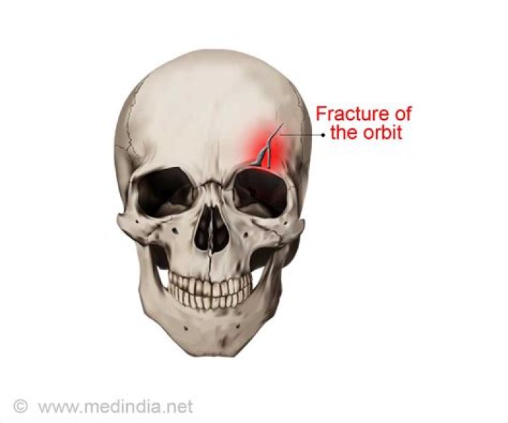
What does a broken eye socket look like
By Victoria Simmons
double vision or reduced vision.swelling of the eyelid.pain, bruising, tearing, or bleeding around the eye.nausea and vomiting (most common in trapdoor fractures)sunken or bulging eye, or droopy eyelid.inability to move your eye in some directions.
How do you know if your eye socket is broken?
- double vision or reduced vision.
- swelling of the eyelid.
- pain, bruising, tearing, or bleeding around the eye.
- nausea and vomiting (most common in trapdoor fractures)
- sunken or bulging eye, or droopy eyelid.
- inability to move your eye in some directions.
What does an eye socket look like?
Your eye socket will look red but it will become pinker in colour as it heals. Some bruising and swelling of the eyelids may occur; this can become worse over the first few days before gradually getting better. You will be able to see the clear plastic shell that has been placed in your eye socket.
What do you do for a broken eye socket?
For a small, uncomplicated blowout fracture that does not affect the movement of your eye, your doctor may prescribe ice packs, decongestants and an antibiotic to prevent infection. You may be told to rest for a few days and to avoid blowing your nose while the eye heals.Can you break eye sockets?
If the bones around your eye are hit hard enough, they can break. This is called an orbital fracture. If your eye socket is treated successfully, and the injury to your eye or tissues around your eye was not too severe, you may not have any long-lasting effects from an eye socket fracture.
What is the white part of the eyeball?
The outer layer of the eyeball is a tough, white, opaque membrane called the sclera (the white of the eye). The slight bulge in the sclera at the front of the eye is a clear, thin, dome-shaped tissue called the cornea.
Does a fractured eye socket hurt?
A broken eye socket usually causes intense pain, swelling, and a black eye, which make it easy to diagnose. The eye socket is the bony structure surrounding and protecting the eye. In addition to the eye, it houses all the muscles, nerves, and connective tissues that connect to and move the eye.
What happens when your eye gets pushed in?
A direct blow to the eye can damage the eyeball, the supporting muscles and ligaments, the eyelid, or the bony eye socket (orbit). Symptoms that may mean there is a more serious injury include: Vision changes. Inability to move the eye normally in all directions.What happens after eye enucleation?
After eye removal surgery there will likely be swelling, bruising, and mild discomfort. A pinkish or watery discharge may occur, and the socket may have a scratchy feeling. These aftereffects are normal and should go away in a few days.
Can doctors take your eye out and put it back in?If globe luxation recurs, a doctor may recommend a surgical procedure called lateral tarsorrhaphy. During this procedure, an eye doctor will sew a portion of the eyelid together to prevent the eye from opening too wide and allowing the eyeball to pop out of place again.
Article first time published onCan you pull out your eye?
For many, the idea of removing an eye can be understandably very daunting. However, it is important to know that eye removal is a relatively common surgery that can treat certain eye diseases, alleviate eye pain, and greatly improve the patient’s quality of life.
Can you take an eye out and put it back in?
You should be able to get your eye back in place without serious, long-term damage. (If the ocular muscles tear or if the optic nerve is severed, your outlook won’t be as clear.)
Is an orbital fracture a medical emergency?
Orbital fractures are a common, potentially vision-threatening presentation to an emergency department. Appropriate early management and referral by the emergency medicine practitioner has a significant role in preventing cosmetic and functional sequelae of orbital trauma.
When should you go to the ER for an eye injury?
We recommend seeking immediate medical attention anytime you experience swelling, redness, or pain in your eye, especially if it occurs after an injury or having a foreign object or chemical in your eye. When left untreated, these injuries can damage your eye even more, leading to partial and/or permanent blindness.
What does it mean when the white part of your eye is blue?
Blue sclera. Blue sclera is caused by a congenitally thinner-than-normal sclera or a thinning of the sclera from disease, which allows the color of the underlying choroidal tissue to show through it.
What are retinas?
The retina is a layer of tissue in the back of your eye that senses light and sends images to your brain. In the center of this nerve tissue is the macula. It provides the sharp, central vision needed for reading, driving and seeing fine detail. Retinal disorders affect this vital tissue.
What is a Chemosis?
Chemosis is swelling of the tissue that lines the eyelids and surface of the eye (conjunctiva). Chemosis is swelling of the eye surface membranes because of accumulation of fluid.
What is the difference between enucleation and Marsupialization?
Enucleation is defined as a complete removal of the cystic lining with healing by primary intention while marsupialization is synonymous with Partsch’s operation17, and is the conversion of a cyst into a pouch1, it requires considerable aftercare and patient cooperation in keeping the cavity clean whilst it resolves …
How long is recovery after eye removal?
Returning to Work: You will be able to return to normal activity soon after surgery. The orbit should heal quickly and you should be able to return to school or work within 2 to 6 weeks. You should not lift more than 10 pounds, strain, or rub your eye for at least 14 days after surgery.
How do you clean an eye socket?
Take the shell out of your eye socket, wash it in soapy water, rinse it well and put it back. It’s possible for the shell to fall out but this is very rare. If it does fall out, clean it as you normally would and put it back in place.
How many pounds of pressure does it take to pop out an eye?
For dynamic loading, the average porcine rupture pressure was 1.64 ± 0.32 MPa, and the average rupture pressure for human eyes was 0.91 ± 0.29 MPa. Significant differences are found between average rupture pressures from all four groups of tests (p = 0.01).
Can you still drive with one eye?
Having vision in just one eye is called monocular vision, and is actually perfectly legal for driving.
Can you bruise your orbital bone?
Orbital fractures are also frequently accompanied by bruising and swelling: a “black eye” or a “shiner”. Of course, bruising and swelling can also occur from more minor injuries to the eye area.
What is a maxillary fracture?
A maxilla fracture happens when the maxilla becomes cracked or broken. This often happens due to injuries to the face, such as from falling, a car accident, getting punched, or running into an object. These injuries can be significant.
How does orbital fracture happen?
Orbital rim fracture — These are caused by a direct impact to the face, most commonly by an automobile dashboard or steering wheel during a car crash. Because a great deal of force is required to cause these fractures, they often occur with extensive injuries to other facial bones, and sometimes injuries to the brain.
What is considered an eye emergency?
An eye emergency is defined as a condition requiring prompt medical attention due to a sudden change in ocular health or vision. Eye trauma, foreign objects in the eye, chemical exposure to the eyes, and ocular infections are all considered eye emergencies and should be treated immediately.
How do you know if eye pain is serious?
- It is unusually severe or accompanied by headache, fever or unusual sensitivity to light.
- Your vision changes suddenly.
- You also experience nausea or vomiting.
- It is caused by a foreign object or chemical splashed in your eye.
What is left retinal detachment?
Retinal detachment describes an emergency situation in which a critical layer of tissue (the retina) at the back of the eye pulls away from the layer of blood vessels that provides it with oxygen and nutrients. Retinal detachment is often accompanied by flashes and floaters in your vision.