What is a gradient in MRI
By Victoria Simmons
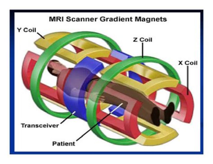
Gradients are simply loops of wire or thin conductive sheets on a cylindrical shell that lies just inside the bore of an MRI Scanner. When an electrical current passes through these coils, the result is a secondary magnetic field. This gradient field distorts the main magnetic field in a slight but predictable pattern.
What is gradient duty cycle in MRI?
An additional gradient parameter commonly measured is duty cycle. The duty cycle does not have a fixed value, but varies by pulse sequence. It represents the percent of time the gradient is able to work at maximum amplitude during that sequence.
What is gradient amplitude?
Sensitivity and peak amplitude The peak amplitude (G) is the strength of the gradient system, i.e. the steepness of the magnetic field. It is measured in mT/m. The gradient amplitude (in combination with the RF excitation pulse) defines the slice thickness.
Why do we need a gradient magnetic field in MRI measurements?
Magnetic Field Gradient If each of the regions of spin was to experience a unique magnetic field we would be able to image their positions. A gradient in the magnetic field is what will allow us to accomplish this.What is the gradient rise time?
The slew rate (AKA “rise time”) is the speed at which the gradient reaches its maximum amplitude. Slew rate is measured in millitesla per meter per microsecond (mT/m/ms). The higher the slew rate, the thinner the anatomical slice, which means higher clarity in the image produced.
What does a magnetic field gradient do?
Magnetic Field Gradients, Spatial Frequencies and k-space As magnetic field gradients establish different precession frequencies across the imaging volume, evolution in a gradient over a specified time creates a spatial distribution of magnetization that varies along the gradient direction.
Which gradient specification is generally the most important when assessing how well an MR system is capable of performing rapid high resolution imaging?
Which gradient specification is generally the most important when assessing how well an MR system is capable of performing rapid, high-resolution imaging? Peak gradient strength. Slew rate.
How do you find the gradient of a magnetic field?
The force/gradient relationship is represented by the formula, F = – ∇ P. Force equation Where F is the force, ∇ is the gradient as a vector quantity and P is the magnetic potential.How many gradient magnetic fields are used for MRI investigation?
Three sets of gradient coils are used in nearly all MR systems: the x-, y-, and z-gradients. Each coil set is driven by an independent power amplifier and creates a gradient field whose z-component varies linearly along the x-, y-, and z-directions, respectively.
What happens in a gradient coil that determines the amplitude of a gradient?The strength/amplitude of the gradient is determined by the amount of current applied to the gradient coil (maximum amplitude determines maximum achievable resolution). Polarity determines which end of the gradient is positive and which is negative.
Article first time published onWhat is spatial gradient field?
The term “spatial gradient magnetic field” refers to the rate at which the static magnetic field strength changes over space or distance per unit of length. This parameter is indicated as dB/dx, using the units of T/m or gauss/cm.
What is peripheral nerve stimulation MRI?
Rapidly changing magnetic fields of gradient coils can induce electric fields in human tissue causing stimulation of peripheral nerves. Magnetic gradient coils are necessary component of every MRI system used to spatial encode the MRI signal.
What affects MRI SAR?
SAR is proportional to the square of the RF frequency (ω). So, all other things being equal, SAR depends on the square of Bo. The same pulse sequence in the same patient imaged at 1.5T would therefore generate 4x the SAR if performed at 3.0T.
What are eddy currents MRI?
Eddy currents (also known as Foucault currents) are the result of rapidly changing gradient magnetic fields that in turn induce stray currents in the surrounding conducting materials. They form in accordance to Faraday’s Law of Induction.
What does the frequency encoding gradient do?
Frequency encoding is used to determine one axis in the xy-plane of the slice. Just like with slice selection, remember that the presence of a magnetic field gradient creates a corresponding gradient in the precession frequencies of the protons along that direction.
How do RF coils work?
Radiofrequency coils (RF coils) are the receivers, and sometimes also the transmitters, of radiofrequency (RF) signals in equipment used in magnetic resonance imaging (MRI). … The receiver coil picks up the oscillations at RF frequencies produced by precession of the magnetic moment of nuclei inside the subject.
What is the unit of strength of magnetic field?
Usually, magnetic field strength is defined by the unit of Oe・A/m ( Oersted・Ampere/meter ). And when it is defined by flux density, the units of G (Gauss) or T (Tesla) are used.
What determines slice thickness in MRI?
The thickness of the slice is determined by a combination of two factors: (1) the strength, or steepness, of the gradient, and (2) the range of frequencies, or bandwidth, in the RF pulse. In most clinical applications, it is desirable to have a series of images (slices) covering a specific anatomical region.
Where do you find frequent use of gradient?
Que.Where do you find frequent use of Gradient?b.MRI Imagingc.PET Scand.None of the mentionedAnswer:Industrial inspection
What are MRI gradient coils made of?
There are three gradient coils in a typical MRI system. They are the resistant-type of electromagnetic coils made from copper or aluminium. They have shields to prevent eddy current interference.
What's better CT or MRI?
Both MRIs and CT scans can view internal body structures. However, a CT scan is faster and can provide pictures of tissues, organs, and skeletal structure. An MRI is highly adept at capturing images that help doctors determine if there are abnormal tissues within the body. MRIs are more detailed in their images.
What is a high gradient magnetic field?
High magnetic field gradient is a crucial factor in HGMS process which can be described as a separation process or a deep-bed filtration process in which a magnetic matrix is magnetized and used to bundle the external magnetic field in its vicinity to generate high magnetic field gradient [12].
How a high gradient magnetic field could affect cell life?
The magnetic gradient forces localized near the MNPs affect cell functions in two main ways: i) changing the resting membrane potential, as predicted by Eq. 1, and ii) generating local magnetic pressure that can cause membrane deformation, resulting in cell membrane blebbing.
Is an MRI a static magnetic field?
In a clinical setting, MRI scanners routinely use static magnetic fields in the range of 200–3000 mT. … In medical research, fields of up to 9400 mT are used to scan the entire body of patients.
What happens in k space when the phase FOV is doubled?
What happens in k-space when the phase FOV is doubled? The incremental step between each line is halved. According to the Nyquist theorem, to avoid aliasing: how many samples are required?
What are the units of k space?
The wavenumber (k) is therefore the number of waves or cycles per unit distance. Since the wavelength is measured in units of distance, the units for wavenumber are (1/distance), such as 1/m, 1/cm or 1/mm.
Why are there gradient coils in an MRI scanner?
The main function of a gradient coil is to spatially modulate the main magnetic field in a predictable way, thereby causing the Larmor frequency of spins to vary as a function of position. This allows spatial encoding of the MR signal.
Where is the spatial gradient magnetic field the greatest?
In general, the highest spatial gradient magnetic field used to assess translational attraction for a medical device is located off-axis, at a side wall, and near the opening of the bore of the scanner [1, 3].
What is an effect associated with the gradient or time varying magnetic fields?
In magnetic resonance, time-varying gradient magnetic fields (dB/dt) may stimulate nerves or muscles by inducing electric fields in patients.
What happens if you twitch during an MRI?
You might feel a twitching sensation during the test. This happens as the MRI stimulates nerves in your body. It’s normal, and nothing to worry about. The MRI scan should take 20-90 minutes.
Where are the peripheral nerves?
Peripheral nerves reside outside your brain and spinal cord. They relay information between your brain and the rest of your body. The peripheral nervous system is divided into two main parts: Autonomic nervous system (ANS): Controls involuntary bodily functions and regulates glands.