
What is Acyanotic heart disease
By Olivia Hensley
Acyanotic heart disease is a heart defect that affects the normal flow of blood. Examples include a hole in the heart wall. The condition is present at birth but may not cause any symptoms or problems until later in life. Sometimes the problem corrects itself during childhood.
What is a Acyanotic heart disease?
Acyanotic heart disease is a heart defect that affects the normal flow of blood. Examples include a hole in the heart wall. The condition is present at birth but may not cause any symptoms or problems until later in life. Sometimes the problem corrects itself during childhood.
What are the major differences of cyanotic heart disease from Acyanotic heart disease?
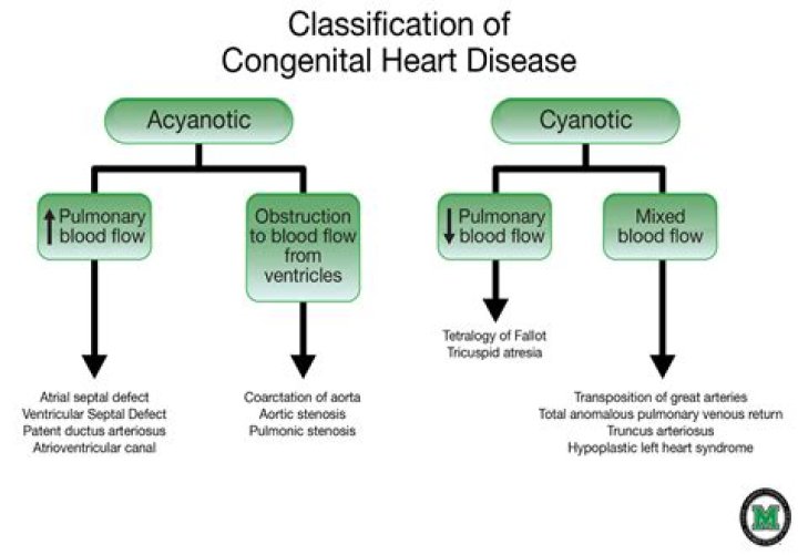
The main difference is that cyanotic congenital heart disease causes low levels of oxygen in the blood, and acyanotic congenital heart disease doesn’t. Babies with reduced oxygen levels may experience breathlessness and a bluish tint to their skin.
What are the diseases in the Acyanotic category?
Acyanotic heart defects include ventricular septal defect (VSD), atrial septal defect (ASD), patent ductus arteriosus (PDA), pulmonary valve stenosis, aortic valve stenosis, and coarctation of the aorta.How is cyanotic heart disease different?
Some useful clues can be employed to differentiate cyanosis of cardiac and pulmonary origins (Table 1). In patients with cardiac cause of cyanosis, respiration is relatively comfortable despite cyanosis, which may worsen on crying. A heart murmur and abnormal cardiac silhouette indicate cardiac defects.
Is Avsd cyanotic or Acyanotic?
Infants with complete atrioventricular septal defect often have a bluish discoloration of the skin and mucous membranes (cyanosis) due to insufficient oxygen supply to these tissues.
Why is it called Acyanotic heart disease?
There are many types of congenital heart defects. If the defect lowers the amount of oxygen in the body, it is called cyanotic. If the defect doesn’t affect oxygen in the body, it is called acyanotic.
Is coarctation of the aorta Acyanotic heart disease?
Coarctation of the aorta is often considered a critical congenital heart defect (critical CHD) because if the narrowing is severe enough and it is not diagnosed, the baby may have serious problems soon after birth. CCHDs also can be detected with newborn pulse oximetry screening.How many types of Acyanotic heart defects are present?
18 Types of Congenital Heart Defects.
Which congenital heart defects are Acyanotic in nature?- Ventricular septal defect (VSD).
- Atrial septal defect (ASD).
- Atrioventricular septal defect.
- Patent ductus arteriosus (PDA).
- Pulmonary valve stenosis.
- Aortic valve stenosis.
- Coarctation of the aorta.
What happens in coarctation of aorta?
With coarctation of the aorta, the lower left heart chamber (left ventricle) of your heart works harder to pump blood through the narrowed aorta, and blood pressure increases in the left ventricle. This may cause the wall of the left ventricle to thicken (hypertrophy).
What is the most common form of cyanotic heart disease?
Tetralogy of Fallot is the most common form of cyanotic congenital heart disease. Cyanosis is the abnormal bluish discoloration of the skin that occurs because of low levels of circulating oxygen in the blood.
Which congenital heart defect is described as the incomplete fusion of the endocardial cushions?
A partial or incomplete atrioventricular septal defect is one in which the part of the ventricular septum formed by the endocardial cushions has filled in, either by tissue from the AV valves or directly from the endocardial cushion tissue, and the tricuspid and mitral valves are divided into two distinct valves.
How is cyanotic heart disease diagnosed?
- Electrocardiogram (ECG). This painless test records the electrical signals in your heart. …
- Chest X-ray. …
- Pulse oximetry. …
- Echocardiogram. …
- Transesophageal echocardiogram. …
- Cardiac CT scan and MRI . …
- Cardiac catheterization.
How common is cyanotic heart disease?
Congenital heart disease (CHD) affects 8 to 9 per 1000 live births, and approximately 25% are considered CCHD. The incidence of CHD increase to 2% to 6% for a second pregnancy after the birth of a child with CHD or if a parent is affected. Tetralogy of Fallot (TOF) is the most common CCHD (5% of all CCHD).
Is coarctation of the aorta left to right shunt?
VSD is frequently present, and coarctation exacerbates the associated left-to-right shunt. Other levels of left heart obstruction (aortic stenosis, subaortic stenosis) may be present and may add to LV afterload.
Is aortic stenosis cyanotic or Acyanotic?
The most common acyanotic lesions are ventricular septal defect, atrial septal defect, atrioventricular canal, pulmonary stenosis, patent ductus arteriosus, aortic stenosis and coarctation of the aorta. In infants with cyanotic defects, the primary concern is hypoxia.
Why is Tetralogy of Fallot cyanotic?
The cause of cyanosis is a lower than normal blood oxygen level. Patients with tetralogy of Fallot are at risk for cyanosis because the narrowing of blood flow to the lungs in combination with a VSD or hole allows blood in many instances to bypass the lungs and go directly up to the body.
Why is Acyanotic left to right?
With physiologic declines in pulmonary vascular resistance, compensatory in utero right ventricular hypertrophy regresses, resulting in a more compliant right ventricle and atrium. This allows a progressive left-to-right increase in ASD shunt volume that is further pronounced with larger defect size.
Is tricuspid atresia cyanotic or Acyanotic?
Tricuspid atresia is the third most common form of cyanotic congenital heart disease, with a prevalence of 0.3-3.7% in patients with congenital heart disease. The deformity consists of a complete lack of formation of the tricuspid valve with absence of direct connection between the right atrium and right ventricle.
Can adults have Tetralogy of Fallot?
Tetralogy of Fallot (ToF) is the most common complex lesion seen in adults with congenital heart disease (CHD). The condition usually is diagnosed at birth or shortly thereafter in response to its hallmark cyanosis.
Is a PFO considered a congenital heart defect?
Patent foramen ovale (PFO) and atrial septal defect (ASD) are congenital (present-at-birth) conditions that affect the inter-atrial septum (tissue between the right and left upper chamber of the heart).
What is the most common congenital heart disease in adults?
Ventricular septal defect (VSD) (see Figures 2 and 3) is the most common congenital heart defect.
Can you live without an aorta?
The consequences can be deadly. As many as 40 percent of people who experience aortic dissection die almost instantly, and the risk of death increases by 3-4 percent every hour the condition is left untreated.
How long can you live with coarctation of the aorta?
Individuals with coarctation of the aorta have historically had poor long-term out- comes with a mean life expectancy of 35 years. Natural history studies demon- strated 90% of individuals dying before age 50 years.
Why do you keep PDA open in coarctation of aorta?
An open (patent) ductus arteriosus is necessary to allow blood flow to the lower part of the body. The ascending aorta supplies blood to the head and arms (depending on the type of interrupted aortic arch), while the lower body receives blood that would otherwise pass through to the lungs.
What do you mean by Tetralogy of Fallot?
Tetralogy of Fallot (pronounced te-tral-uh-jee of Fal-oh) is a birth defect that affects normal blood flow through the heart. It happens when a baby’s heart does not form correctly as the baby grows and develops in the mother’s womb during pregnancy.
How is Avsd repaired?
Open-heart surgery is the mainstay of treatment for children with AVSD. The repair involves placement of one or two patches to divide the common valve into right and left sides and close the holes. This is performed after beginning heart/lung bypass to support the circulation during the repair itself.
Can you have Avsd without Down syndrome?
In patients with AVSD without Down syndrome, about 20% have other genetic disorders such as Holt-Oram or Noonan syndrome.
What is heart IVS?
The interventricular septum (IVS, or ventricular septum, or during development septum inferius) is the stout wall separating the ventricles, the lower chambers of the heart, from one another.
How do you fix cyanosis?
- Warming of the affected areas. …
- Surgery as a treatment for cyanosis. …
- Oxygenation as a treatment for cyanosis. …
- Intravenous fluids. …
- Drugs as a treatment for cyanosis. …
- Immunizations for children with cyanosis. …
- Injections for babies with cyanosis. …
- Glucose administration.