What is head and neck anatomy
By Olivia Bennett
In radiology, the ‘head and neck’ refers to all the anatomical structures in this region excluding the central nervous system, that is, the brain and spinal cord and their associated vascular structures and encasing membranes i.e. the meninges.
What is in the neck anatomy?
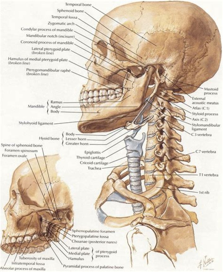
Some important structures contained in or passing through the neck include the seven cervical vertebrae and enclosed spinal cord, the jugular veins and carotid arteries, part of the esophagus, the larynx and vocal cords, and the sternocleidomastoid and hyoid muscles in front and the trapezius and other nuchal muscles …
What is the neck anatomy called?
The neck region of the spine is known as the Cervical Spine. This region consists of seven vertebrae, which are abbreviated C1 through C7 (top to bottom). These vertebrae protect the brain stem and the spinal cord, support the skull, and allow for a wide range of head movement.
What Is a head in anatomy?
head, in human anatomy, the upper portion of the body, consisting of the skull with its coverings and contents, including the lower jaw. … The term also is used to describe the anterior or fore part of animals other than humans.What is the function of the head and neck?
The bones of the head and neck play the vital role of supporting the brain, sensory organs, nerves, and blood vessels of the head and protecting these structures from mechanical damage. Movements of these bones by the attached muscles of the head provide for facial expressions, eating, speech, and head movement.
What parts are located in the head?
- A head is the part of an organism which usually includes the ears, brain, forehead, cheeks, chin, eyes, nose, and mouth, each of which aid in various sensory functions such as sight, hearing, smell, and taste, respectively. …
- Heads develop in animals by an evolutionary trend known as cephalization.
What connects the head and neck?
The occipital bone is the only bone in your head that connects with your cervical spine (neck). The occipital bone surrounds a large opening known as the foramen magnum. The foramen magnum allows key nerves and vascular structures passage between the brain and spine.
What is side of head called?
The temple is a juncture where four skull bones fuse together: the frontal, parietal, temporal, and sphenoid. It is located on the side of the head behind the eye between the forehead and the ear.What is the neck?
The neck is the part of the body that separates the head from the torso. The Latin-derived term cervical means “of the neck.” The neck supports the weight of the head and is highly flexible, allowing the head to turn and flex in different directions.
What does neck mean in medical terms?The cervical spine, also known as the neck, is comprised of seven vertebral bodies (C1-C7) that make up the upper most part of the spine.
Article first time published onWhere is C4 5 and C5 6?
The C4 and C5 vertebrae are the primary members of the mid-cervical spine. These two members are the most mobile in the mid-neck, and they support most of the forward and backward movements of this section of the neck.
Where is C6 and C7?
The C6 C7 spinal motion segment is located in the lower part of the cervical spine and consists of the C6 and C7 vertebrae, and the anatomical structures connecting them. This segment helps provide neck flexibility, supports the cervical spine and head, and protects the spinal cord and nerve pathways.
What's the back of the neck called?
The cervical spine – the neck and upper back, composed of the seven vertebrae closest to the skull. The cervical spine supports the weight and movement of your head and protects the nerves exiting your brain.
What are the 4 basic functions of the head and neck muscles?
- swallowing and chewing (mastication)
- making facial expressions.
- moving your head and neck.
- supporting your head.
What is the neck function?
Its primary function is to provide support for the skull, while still allowing for movement. It is the most flexible part of the spine. This flexibility allows for large movements to scan our surroundings. The majority of sensory inputs occur at the head; thus, proper neck movement is vital to our survival.
How does the neck work?
Many in the neck help to stabilize or move the head. Some also create facial expressions. The fan-shaped trapezius muscles extend from the back of the skull down to the middle of the back, along the spine, and fan over into the shoulders. These muscles give the sides of the neck their shape.
Is head and neck anatomy hard?
Abstract : Statement of the Problem: The head and neck structures are for learning probably the most difficult part of anatomy. The main problem is that most structures of the head and neck are too small and located deeply, in hardly accessible areas.
Which includes the head neck and trunk?
Included in the axial skeleton are the head, vertebral column, sternum, and ribs.
What is atlas and Axis?
The atlas and axis vertebrae are the two most superior bones in the vertebral column, and they are part of the seven cervical vertebrae. The atlas is the top-most bone, sitting just below the skull; it is followed by the axis. Together, they support the skull, facilitate neck movement, and protect the spinal cord.
Where is the head located?
In human anatomy, the head is at the top of the human body. It supports the face and is maintained by the skull, which itself encloses the brain.
What are the two parts of the head?
The skull is composed of two parts: the cranium and the mandible. In humans, these two parts are the neurocranium and the viscerocranium (facial skeleton) that includes the mandible as its largest bone.
How many parts are in the head?
The adult human skull consists of two regions of different embryological origins: the neurocranium and the viscerocranium. The neurocranium is a protective shell surrounding the brain and brain stem. The viscerocranium (or facial skeleton) is formed by the bones supporting the face.
Is throat part of neck?
In vertebrate anatomy, the throat is the front part of the neck, internally positioned in front of the vertebrae. It contains the pharynx and larynx. An important section of it is the epiglottis, separating the esophagus from the trachea (windpipe), preventing food and drinks being inhaled into the lungs.
Where is the weakest part of the skull?
The pterion is known as the weakest part of the skull. The anterior division of the middle meningeal artery runs underneath the pterion. Consequently, a traumatic blow to the pterion may rupture the middle meningeal artery causing an epidural haematoma.
Where is the crown of your head located?
The crown of your head is located at the very top of your skull. You may also sometimes see it referred to as the vertex. Like other parts of your skull, the crown works to provide protection and support for the tissues of your head, including your brain.
What is top of skull called?
This cavity is bounded superiorly by the rounded top of the skull, which is called the calvaria (skullcap), and the lateral and posterior sides of the skull. The bones that form the top and sides of the brain case are usually referred to as the “flat” bones of the skull.
What are symptoms of nerve damage in neck?
- A sharp pain in the arm.
- Pain in the shoulder.
- A feeling of numbness or pins and needles in the arm.
- Weakness of the arm.
- Worsening pain when you move your neck or turn your head.
Where is C5 C6 and C7?
The C5-C6 spinal motion segment (located in the lower cervical spine just above the C7 vertebra) provides flexibility and support to much of the neck and the head above.
What does C7 nerve affect?
C7 helps control the triceps (the large muscle on the back of the arm that straightens the elbow) and wrist extensor muscles. The C7 dermatome goes down the back of the arm and into the middle finger. C8 helps control the hands, such as finger flexion (handgrip).
What causes C7 to stick out?
The vertebra located at the base of your neck, the cervical C7 vertebrae is also called the first thoracic vertebrae. It’s the one that feels like it sticks out when you run your hand down the back of your neck. It’s directly associated, when out of alignment, with issues like shoulder and elbow bursitis.
What activities should be avoided with cervical spinal stenosis?
- Avoid stretching in a standing position and extension stretches. …
- Instead, try stretching while laying down. …
- Avoid doing free weights. …
- Instead, try using a weight machine. …
- Avoid running and similar high-impact exercises. …
- Instead, try swimming, cycling, or an elliptical machine.