What is meant by the visual pathway where is the blind spot and what causes it
By Christopher Green
blind spot, small portion of the visual field of each eye that corresponds to the position of the optic disk
What is meant by the visual pathway?
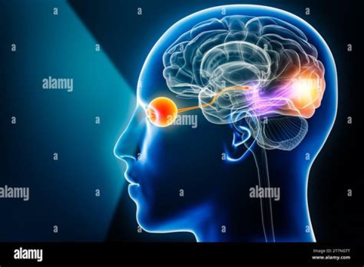
The visual pathway is the pathway over which a visual sensation is transmitted from the retina to the brain. This includes a cornea and lens that focuses images on the retina, and nerve fibers that carry the visual sensations from the retina through the optic nerve.
What is blind spot and where it is located?
The blind spot is the location on the retina known as the optic disk where the optic nerve fiber exit the back of the eye.
What is a blind spot and what causes it?
What causes a blind spot in the eye? Each of our eyes has a tiny functional blind spot about the size of a pinhead. In this tiny area, where the optic nerve passes through the surface of the retina, there are no photoreceptors. Since there are no photoreceptor cells detecting light, it creates a blind spot.What is the visual pathway and where?
The visual pathway begins with photoreceptors in the retina and ends in the visual cortex of the occipital lobe. The photoreceptors are cells of two types: rods and cones. Rods play a special role in peripheral vision and in vision under low light conditions.
What is the Where pathway responsible for?
The dorsal stream (or, “where pathway”) leads to the parietal lobe, which is involved with processing the object’s spatial location relative to the viewer and with speech repetition.
Where does the visual pathway begin?
The optic pathway begins in the retina, which is a complex structure made up of ten different layers. Each layer serves a distinct function. The photoreceptor layers consist of the rods and cones, which generate action potentials with the help of rhodopsin through photosensitive cycles.
Where is the blind spot located when driving?
Blind spots are the areas to the sides of your car that can’t be seen in your rear mirror or side mirrors– to make sure these spots are clear before changing lanes, you’ll have to physically turn around and look to see what kind of crazy stuff is going on out there.What do blind spots in vision mean?
Blind spots are sometimes linked to problems like migraines, glaucoma, retinal detachment, macular degeneration, diabetic retinopathy, and HIV/AIDS-related eye problems. Talk to your doctor if: You see blank or dark spots in your field of vision. You notice a blind spot when you’re doing everyday activities.
What is blind spot Class 10?Blind spot is the region where the optic nerve passes through the optic disk and out of the eyes. Also, it is at this very region that the blood vessels enter the eyes. It lacks photoreceptor cells (rods and cones) in the retina so the light falling at this spot does not form any image.
Article first time published onWhat is the blind spot in the eye and how does it impact the transduction of light energy?
The eye’s retina receives and reacts to incoming light and sends signals to the brain, allowing you to see. One part of the retina, however, doesn’t give you visual information—this is your eye’s “blind spot.”
Why do we have a blind spot in each eye quizlet?
Why do we have a blind spot, and what are two reasons we do not perceive a dark patch in our visual field? There are no photoreceptors on the optic disc, therefore we have a blind spot. We don’t see a big hole because we have 2 eyes so whatever one eye doesn’t catch the other one will.
What is the visual pathway from the eyes to the occipital lobe?
The visual pathways comprise the optic nerve, optic chiasm, optic tract, optic radiation and the visual cortex in the occipital lobes. Nerve impulses arising in the retina travel via the optic nerve to the optic chiasm.
Where is the dorsal visual pathway?
Dorsal visual pathway: this pathway extends from the primary visual cortex (V1) in the occipital lobe to the parietal lobe. The dorsal pathway is subdivided by the intraparietal sulcus (IPS) into several main sectors including the superior parietal lobule, inferior parietal lobule, and the supramarginal gyrus.
What are the steps of the visual pathway?
- Light enters the eye through the cornea. …
- From the cornea, the light passes through the pupil. …
- From there, it then hits the lens. …
- Next, light passes through the vitreous humor. …
- Finally, the light reaches the retina.
Where is the optic pathway?
The optic tract is an extension of the optic nerve located in the brain. It begins at the area where information from the left eye and right eye cross (or “decussate”) to create a complete visual picture. The optic tract is actually comprised of two separate tracts: the left optic tract and the right optic tract.
How is the visual pathway from the eye different from that of the ear?
How is the visual pathway from the eye different from that of the ear or hand? Each eye is not primarily connected to one hemisphere only. Briefly explain split-brain research.
What is the pathway of the optic nerve?
The optic pathway includes the retina, optic nerve, optic chiasm, optic radiations, and occipital cortex (see figure Higher visual pathways. Damage along the optic pathway causes a variety… read more ). Damage along the optic pathway causes a variety of visual field defects.
What is the pathway of light through the eye and to the visual cortex?
The projection from the LGN to the visual cortex is called the optic radiation. Because damage at any point along the pathway from the retina to the cortex results in some degree of blindness, this is clearly the pathway through which conscious visual perception takes place in human beings.
What is the secondary visual pathway?
Tutorial 24: Brain Visual Pathways. … This secondary pathway mediates the ability to localize visual objects in space. In addition, along this pathway, visual information is integrated with auditory and somatosensory information and used to control movements made to objects of interest.
Where are your blind spot quizlet?
what is a blind spot? they are areas toward the side and rear of your vehicle that you cannot see with your mirrors or side vision.
Where are blind spot sensors located?
The blind spot detection system uses the radar sensors in the rear of the vehicle. These sensors are normally behind the rear bumper on each side. However, there are some instances where the sensor is in a different location, such as in the tail light or in the quarter panel behind the bumper cover.
What is blind spot in the eye Class 8?
Blind spot is a small area of the retina insensitive to light where the optic nerve leaves the eye . When the image of an object is formed at the blind spot in the eye, it cannot be seen by the eye. Blind spot is not sensitive to light because there are no light-sensitive cells like rods or cones in this region.
What is blind spot Class 12?
Complete answer: Blind spot is the small portion present in the eye. It is present where the optic nerves leave the retina of the eye. There are no photoreceptors present in this area making it a region insensitive to the light.
What is the blind spot Class 8?
A small portion in the retina which is insensitive to light is called blind spot. Blind spot is an area on the retina where the nerve endings enter the optic nerves. Since, this area has no visual receptors such as rods and cones, the images falling on this area cannot be detected.
Where are rods and cones located?
The retina of the eye has two types of light-sensitive cells called rods and cones, both found in layer at the back of your eye which processes images.
What structures exit enter the eye at the blind spot?
The optic disc or optic nerve head is the point of exit for ganglion cell axons leaving the eye. Because there are no rods or cones overlying the optic disc, it corresponds to a small blind spot in each eye. The ganglion cell axons form the optic nerve after they leave the eye.
What is the difference between the fovea and the blind spot?
Visual acuity such as sharpness and detail is greatest at the fovea, while at the blind spot it is insensitive to visual stimulation, it’s the part of the retina that converges to the optic nerve.
Where does the visual pathway cross so that each optic tract carries impulses from the opposite visual field?
The visual pathway The fibres from the nasal halves of each retina cross to the opposite side of the brain, while those from the temporal halves remain uncrossed. This partial decussation is called the chiasma. The optic nerves after this point are called the optic tracts, containing nerve fibres from both retinas.
How visual information is transmitted to the brain?
The optic nerve, a cable–like grouping of nerve fibers, connects and transmits visual information from the eye to the brain. The optic nerve is mainly composed of retinal ganglion cell (RGC) axons. … having a long axon that extends into the brain via the optic chiasm and the optic tract. synapsing with the LGN.
Where visual images are perceived?
Visual perception takes place in the cerebral cortex and the electrochemical signal travels through the optic nerve and via the thalamus (another area of the brain) to the cerebral cortex.