What is Quadranopsia
By Olivia Bennett
quadrantanopia. A loss of vision in a quarter of the visual field. The defect is usually bilateral as it is typically caused by a lesion past the optic chiasma.
Can quadrantanopia be cured?
Any field loss present after this time may be permanent. You may however feel that your sight improves as you adapt to the defect over time. Visual field loss cannot be cured if it does not spontaneously recover.
What causes homonymous superior quadrantanopia?
Homonymous superior quadrantanopia is caused by damage to the contralateral inferior parts of the posterior visual pathway: the inferior optic radiation (temporal Meyer loop), or the inferior part of the occipital visual cortex below the calcarine fissure.
Can a stroke cause quadrantanopia?
The presence of sudden onset bilateral homonymous quadrantanopia is a red flag for stroke in the occipital cortex or the optic radiation.What lesion causes quadrantanopia?
Quadrantanopia, quadrantanopsia, refers to an anopia affecting a quarter of the field of vision. It can be associated with a lesion of an optic radiation. While quadrantanopia can be caused by lesions in the temporal and parietal lobes, it is most commonly associated with lesions in the occipital lobe.
What does Quadrantanopia look like?
Quadrantanopia describes defects confined mostly to about one fourth of an eye’s visual space. Homonymous describes defects that affect the same side of the vertical meridian (i.e., right or left side) of both eyes.
Can I drive with quadrantanopia?
In addition, there should be no significant defect in the binocular field that encroaches within 20° of the fixation above or below the horizontal meridian. This means that homonymous or bitemporal defects that come close to fixation, whether hemianopic or quadrantanopic, are not usually acceptable for driving.
What causes inferior Quadrantanopia?
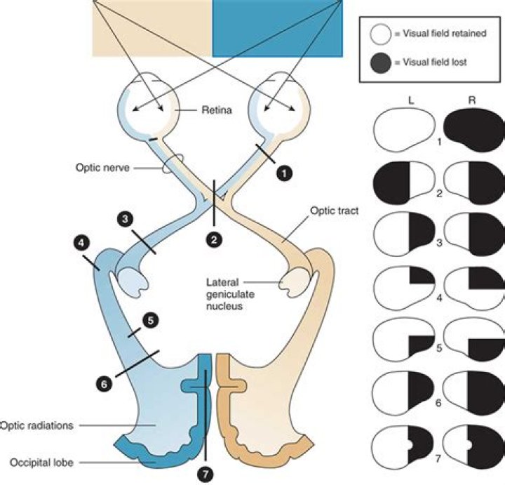
A superior quadrantanopia results from an insult to the optic radiation inferiorly in the temporal lobe, resulting in a ‘pie in the sky’ type of visual field defect (Figure 1d), while an inferior quadrantanopia is caused by damage to the parietal lobe optic radiation (Figure 1e).What do stroke victims see?
Examples in stroke survivors include rapid eye jiggling (nystagmus), eye turning (strabismus), eye tracking control issues (oculomotor dysfunction) and double vision (diplopia). Your depth perception, balance, coordination and overall vision may be affected by these.
What part of the brain causes hemianopia?As for the areas of the brain most affected, 40% of homonymous hemianopsias originate in the occipital (rear) lobe of the cerebral hemisphere.
Article first time published onHow can I regain my peripheral vision?
- Why Peripheral Vision Is Important For Athletes. …
- The Toothpick And Straw Method. …
- Use Cognitive Training Gear. …
- Try Recording Things That Are Outside Your Central Field Of View. …
- Do Sports Drills That Challenge Your Peripheral Vision. …
- Aim For A Good Diet And Lifestyle.
What causes pie in the sky defect?
Left Superior Homonymous Quadrantanopia: This visual defect is often referred to as pie in the sky. This visual defect happens when the inferior optic radiating fibers (Meyer’s loop) are damaged in the temporal lobe of the brain. Strokes involving the middle cerebral artery (MCA) can result in this presentation.
What is congruous and incongruous?
A congruous visual field defect is identical between the two eyes, whereas an incongruous defect differs in appearance between the eyes.
What is the ICD 10 code for Quadrantanopia?
ICD-10:H53.461Short Description:Homonymous bilateral field defects, right sideLong Description:Homonymous bilateral field defects, right side
What are the optic chiasm?
Listen to pronunciation. (OP-tik ky-AZ-muh) The place in the brain where some of the optic nerve fibers coming from one eye cross optic nerve fibers from the other eye. Also called optic chiasm.
What causes Binasal hemianopia?
The pathophysiology of binasal hemianopsia, while not well understood, is hypothesized to be associated with damage caused by increased intracranial pressure and/or compression from adjacent arteries such as the internal carotid or anterior cerebral caused by shifting from nearby intracranial tumors.
What is homonymous superior quadrantanopia?
Homonymous superior quadrantanopia, also called “pie in the sky,” causes a field deficit in the superior field of both eyes for the same side. This visual field deficit is bilateral and involves retrochiasmal pathways.
Where are optic radiations?
The optic radiations, or the geniculocalcarine tract, are a projection tract that connects the lateral geniculate nucleus to the primary visual cortex in the occipital lobe.
What happens if you fail DVLA eye test?
Answer: The DVLA will assess the results of your visual field test based on their criteria. If they revoke your driving license, there is an appeal process and the DVLA will inform you of this at the same time. You should seek professional advice if you are concerned about your eye health.
What is the minimum visual field for driving?
Drivers must have a horizontal field of vision of at least 120 degrees. In addition, the extension should be at least 50 degrees left and right and 20 degrees up and down. No defects should be present within the radius of the central 20 degrees. This requirement applies to drivers who are binocular or monocular.
Do you have to have a medical at 70 to drive?
It is a very good idea to have a medical check before renewing your licence when you reach 70 years of age, and again each time your licence is renewed.
Is loss of peripheral vision a disability?
Yes, peripheral vision loss is considered a disability, since the loss of peripheral vision can affect one or both eyes, hindering the interaction of the individual with their surroundings.
What are the 5 warning signs of a stroke?
- Sudden numbness or weakness in the face, arm or leg (especially on one side of the body).
- Sudden confusion or trouble speaking or understanding speech.
- Sudden vision problems in one or both eyes.
- Sudden difficulty walking or dizziness, loss of balance or problems with coordination.
Does double vision go away after a stroke?
About one-third of stroke survivors experience vision loss. Most people who have vision loss after a stroke do not fully recover their vision. Some recovery is possible – this will usually happen in the first few months after a stroke.
Does a stroke affect eyesight?
Stroke can affect the visual pathways of your eye and this can affect your vision in different ways including: visual field loss. blurry vision. double vision.
Why do you get macular sparing in PCA stroke?
Macular sparing may be caused by collateral vascular supply to the macular region or by the very large macular representation in the occipital cortex; additionally, bilateral representation of macular vision has been suspected.
What kind of stroke causes homonymous hemianopia?
Background: Previous reports have suggested that most cases of homonymous hemianopia (HH) are caused by occipital stroke. However, these reports have not always been supported by brain imaging.
How does a stroke cause homonymous hemianopia?
Homonymous hemianopsia occurs because the right half of the brain has visual pathways for the left hemifield of both eyes, and the left half of the brain has visual pathways for the right hemifield of both eyes. When one of these pathways is damaged, the corresponding visual field is lost.
What part of the brain affects the eyes?
Occipital lobe. The occipital lobe is the back part of the brain that is involved with vision.
Can you recover from hemianopia?
Spontaneous recovery of HH In a 15-year longitudinal study, Zhang et al. (2006b) analyzed spontaneous recovery in hemianopia patients. They observed recovery approximately 38.4% of the cases within the commonly accepted period of 6 months (after which, the HH becomes chronic).
How can I improve my hemianopia?
- wearing prismatic correction glasses to help with double vision.
- getting vision compensatory training to help you use your remaining vision more efficiently.
- undergoing vision restoration therapy to improve visual information processing.