What is radiographic testing in NDT
By James Craig
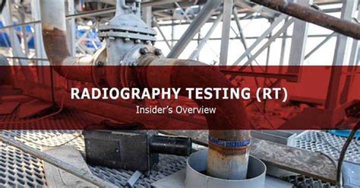
Radiographic Testing (RT) is a non-destructive testing (NDT) method which uses either x-rays
Why is radiography test required?
Radiographic testing provides a permanent record in the form of a radiograph and provides a highly sensitive image of the internal structure of the material. The amount of energy absorbed by the object depends on its thickness and density. Energy not absorbed by the object causes exposure of the radiographic film.
What is radiographic analysis?
It is used to diagnose or treat patients by recording images of the internal structure of the body to assess the presence or absence of disease, foreign objects, and structural damage or anomaly. During a radiographic procedure, an x-ray beam is passed through the body.
Where is radiographic testing used?
Radiographic testing is widely used in a variety of industry sectors including aerospace, power generation, construction, petroleum, chemical and automotive, and for all types of components and parts.What are radiographic films?
X-ray films for general radiography consist of an emulsion-gelatin containing radiation sensitive silver halide crystals, such as silver bromide or silver chloride, and a flexible, transparent, blue-tinted base.
How many types of radiography are there?
There are three types of diagnostic radiographs taken in today’s dental offices — periapical (also known as intraoral or wall-mounted), panoramic, and cephalometric. Periapical radiographs are probably the most familiar, with images of a few teeth at a time captured on small film cards inserted in the mouth.
What is the principal advantage of the radiographic inspection method of NDT?
Following exposure to radiation, the film is then processed and then viewed on an illuminated screen for visual interpretation of the image. Radiography gives a permanent record (the exposed film), which is a major advantage of the method, and is widely used to detect volumetric flaws (surface and internal).
How radiographic images are formed?
A radiographic image is created by passing an x-ray beam through the patient and interacting with an image receptor, such as an imaging plate in computed radiography (CR). The variations in absorption and transmission of the exiting x-ray beam structurally represent the anatomic area of interest.What is radiographic testing in welding?
Radiographic Testing (RT) – This method of weld testing makes use of X-rays, produced by an X-ray tube, or gamma rays, produced by a radioactive isotope. … Energy not absorbed by the object will cause exposure of the radiographic film. These areas will be dark when the film is developed.
What is the difference between radiology and radiography?Radiology encompasses not only imaging techniques, such as x-rays, but also treatments, such as radiation therapy. … Radiography is limited to performing the actual imaging tests. These tests are X-rays, CT scans and MRI procedures.
Article first time published onWhat are the radiographic equipment?
- Radiographic Equipments. Xray rooms vary in design, depending on their purpose. …
- The Xray Tube. The xray tube is the source of radiation. …
- Xray Tube Housing. …
- Xray Tube Support. …
- Collimator. …
- Radiographic Table. …
- Tilting Table. …
- Floating Tabletop.
Is radiography a code test?
ISO 5579:2013(en), Non-destructive testing — Radiographic testing of metallic materials using film and X- or gamma rays — Basic rules.
What is the difference between a radiograph and a film?
In photography, reflected light rays from the object expose the film to produce an image. In radiography, X-rays that pass through the object expose the film to produce an image.
What are the two main parts of radiographic film?
The radiographic film is composed of a base and an emulsion layer joined together by the substratum. The emulsion may be coated on one side (single emulsion film) or both sides (double emulsion film). Intra-oral dental films are coated on both sides as this provides increased film speed.
What are the parameters in radiographic testing?
The major parameters are spatial resolution, contrast sensitivity and optical density range. Derived from the properties of X-ray NDT film systems and application ranges minimum requirements are defined.
What is the disadvantage of radiography test?
Radiography limitations: Relatively expensive equipment. Relatively slow inspection process. Sensitive to flaw orientation. Usualy not possible to determine depth of indications.
What is radiography in oil and gas?
Radiographic Testing (RT) is a nondestructive examination (NDE) technique that involves the use of either x-rays or gamma rays to view the internal structure of a component. In the petrochemical industry, RT is often used to inspect machinery, such as pressure vessels and valves, to detect for flaws.
What is radiology type?
Diagnostic radiology is a medical specialisation that involves undertaking a range of imaging procedures to obtain images of the inside of the body. … Diagnostic radiology is at the core of clinical decision-making in modern medicine. Diagnostic imaging tests can include: X-rays (plain radiography)…
What is difference between radiography and ultrasonic testing?
About industrial radiography and ultrasonic testing Let’s start with radiography. This method uses X-rays and gamma rays that penetrate a solid object (our welds and weld interiors) and then go onto a photographic film. … In contrast to radiography, ultrasonic testing uses mechanical vibrations and ultrasonic waves.
What type of radiation is used in radiography?
X-rays are a form of electromagnetic radiation, similar to visible light. Unlike light, however, x-rays have higher energy and can pass through most objects, including the body. Medical x-rays are used to generate images of tissues and structures inside the body.
What is radiographic image quality?
IMAGE QUALITY. Radiographic Quality Radiographic Quality refers to the fidelity with which the anatomic structures being examined are imaged on the film. Three main factors: Film Factors Geometric Factors Subject Factors.
What is radiographic distortion?
— Distortion may be defined, from a radiographic standpoint, as a variation in the size or shape of an object as shown on the film from its true size or shape. … Magnified distortion is influenced by the distance of the object to be radiographed from the film, and the distance of the focal spot of the tube from the film.
What is radiographic contrast?
Contrast is the difference in density or difference in the degree of grayness between areas of the radiographic image.
What are the branches of radiography?
There are two branches of radiography: Diagnostic and Therapeutic.
Can a radiographer be a radiologist?
Although it can vary, most states require radiographers be licensed or certified. The two professions are different, but they are dependent on each other. Radiologists and radiographers work together. Radiologists depend on radiographers to provide quality scans so they can interpret the results.
How is RT film sensitivity calculated?
As with the wire IQI, the material and dimensions of the step wedge are selected to match the application. The diameter of the smallest hole visible on the radiograph determines the sensitivity, this being calculated as hole diameter divided by component thickness expressed as a percentage.
What is geometric unsharpness?
Geometric unsharpness refers to the loss of definition that is the result of geometric factors of the radiographic equipment and setup. It occurs because the radiation does not originate from a single point but rather over an area. … As the source size decreases, the geometric unsharpness also decreases.
What does the word radiograph mean?
Definition of radiograph (Entry 1 of 2) : a picture produced on a sensitive surface by a form of radiation other than visible light specifically : an X-ray or gamma ray photograph. radiograph.