
What is the interventricular septum
By Victoria Simmons
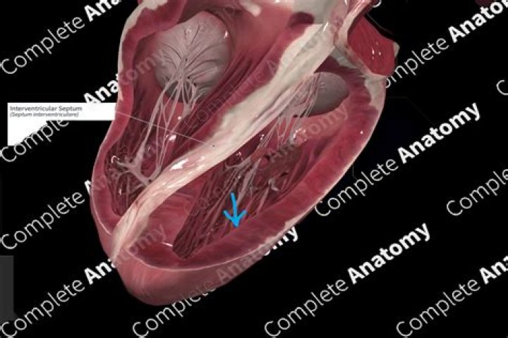
The interventricular septum separates the left ventricle and the right ventricle. It is muscular at the apex and tapers to a membranous portion at the heart base near the origin of the aorta.
What is the function of the interventricular and interatrial septum?
cardiovascular system. …a partition known as the interatrial septum; the lower chambers, the ventricles, are separated by the interventricular septum. The atria receive blood from various parts of the body and pass it into the ventricles. The ventricles, in turn, pump blood to the lungs and to the remainder of the body …
Why is the interventricular septum important?
The interventricular septum separates the left ventricle and the right ventricle. It is muscular at the apex and tapers to a membranous portion at the heart base near the origin of the aorta. Septal defects may occur in any area of the septum, but are most commonly located in the membranous portion.
What are the parts of the interventricular septum?
The right aspect of the muscular ventricular septum can be designated as having 3 components corresponding to the three portions of the ventricle: inlet, apical trabecular, and outlet.What is the difference between the interatrial and interventricular septum?
That portion of the septum that separates the two upper chambers (the right and left atria) of the heart is termed the atrial (or interatrial) septum while the portion of the septum that lies between the two lower chambers (the right and left ventricles) of the heart is called the ventricular (or interventricular) …
Where is the interventricular septum found?
Interventricular Septum | Atlas of Human Cardiac Anatomy. Location: Medial wall of the left ventricle. This is the wall that borders septum between the left and right ventricle.
Why is interventricular septum located in the heart of birds and mammals What is its function?
The interventricular septum is located in the heart of birds and mammals because they have 4-chambered heart, that is, 2 atria and 2 ventricles. This is where the separation of oxygenated and de-oxygenated blood takes place.
Why does the interventricular septum bulge into the right ventricle?
Due to this angulation, the right ventricle tends to lie anteriorly and the left ventricle posteriorly. Throughout most of its surface area, the septum is as muscular as the left ventricle. It tends to bulge into the chamber of the right ventricle producing a concavity on the left ventricular side.What is the function of the anterior interventricular sulcus?
role in cardiovascular system Shallow grooves called the interventricular sulci, containing blood vessels, mark the separation between ventricles on the front and back surfaces of the heart. There are two grooves on the external surface of the heart.
What is the interventricular groove?Interventricular groove may refer to: Anterior interventricular sulcus, one of two grooves that separates the ventricles of the heart, near the left margin. Posterior interventricular sulcus, one of the two grooves that separates the ventricles of the heart, near the right margin.
Article first time published onIs interventricular septum part of myocardium?
Thus, the myocardium, including the interventricular septum, is uniformly expanded during saline infusion and pericardiotomy. The interventricular septum behaves as part of the left ventricle during aortic and pulmonary artery constriction.
What is the structure and function of the septum?
The ventricular septum is a thick structure comprised of discrete muscular bands that separates the left and right ventricles, and contributes to cardiac function.
What is atria and atrium?
The upper two heart chambers are called atria. Atria are separated by an interatrial septum into the left atrium and the right atrium. The lower two chambers of the heart are called ventricles. Atria receive blood returning to the heart from the body and ventricles pump blood from the heart to the body.
Which animal has a complete interventricular septum?
Archosaurs (crocodilians, birds) and mammals independently evolved complete ventricular septation. Birds and mammals have lost either a left (lAo) or right (rAo) aorta.
Which ventricle does the interventricular septum contract with?
The interventricular septum functions as the posterior and left wall of the right ventricle.
What is the difference between interventricular sulcus and interventricular septum?
The anterior interventricular sulcus is a groove located on the anterior part of the heart, while the posterior interventricular sulcus is located on the posterior part of the heart. These two landmarks form the margins of the interventricular septum.
What is the anterior interventricular a called clinically?
The anterior interventricular artery, often clinically termed the left anterior descending artery, is a branch of the left coronary artery. It originates at the left margin of the pulmonary trunk, anterior to the left atrial auricle.
What does anterior interventricular sulcus contain?
The other, the anterior interventricular sulcus, runs along the line between the right and left ventricles and contains a branch of the left coronary artery.
What is the difference between atria and ventricles?
The two atria are thin-walled chambers that receive blood from the veins. The two ventricles are thick-walled chambers that forcefully pump blood out of the heart.
When the RV is in a volume overload state the interventricular septum will flatten to a D shape in?
The higher the RV pressure, the further the septum will displace into the LV resulting in a D shaped LV cavity (fig 1). Of note, septal flattening in the presence of elevated RV pressure should be distinguished from (isolated) RV volume overload, which leads to a septal flattening during diastole.
What is the posterior interventricular sulcus?
The posterior interventricular sulcus or posterior longitudinal sulcus is one of the two grooves that separates the ventricles of the heart and is on the diaphragmatic surface of the heart near the right margin.
What forms the atrioventricular septum?
The atrioventricular canal enlarges to the right, allowing blood flow to both primitive right and left ventricles. The dorsal and ventral endocardial cushions, along with 2 lateral AV cushions, grow and eventually fuse, forming a septum that divides the right and left AV canals.
Why is it called the septum?
In biology, a septum (Latin for something that encloses; plural septa) is a wall, dividing a cavity or structure into smaller ones.
What do ventricles do?
A ventricle is one of two large chambers toward the bottom of the heart that collect and expel blood received from an atrium towards the peripheral beds within the body and lungs. The atrium (an adjacent/upper heart chamber that is smaller than a ventricle) primes the pump.
What are atriums in the heart?
The heart has four chambers: two atria and two ventricles. The right atrium receives oxygen-poor blood from the body and pumps it to the right ventricle. The right ventricle pumps the oxygen-poor blood to the lungs. The left atrium receives oxygen-rich blood from the lungs and pumps it to the left ventricle.
What is another name for atria?
forecourtspiazzascourtyardsspacesquaresareasconcourses
Which is the reptile has well developed interventricular septum?
Adult turtles, lizards and snakes have a complex ventricle with three cava, partially separated by the horizontal and vertical septa.
Do birds have septum between ventricles?
Crocodilians, birds and mammals, however, have completely separated left and right ventricles, a clear example of convergent evolution. … It is homologous to the inlet septum in mammals and birds. Eventually, the various septal components merge to form the completely septated heart.
Why do birds and mammals have a septum between left and right sides of the heart?
The separation of the right and left side of heart is useful to prevent oxygenated blood and deoxygeneted blood from mixing. Such separation allows a highly efficient supply of oxygen to the body.