What is the purpose of arachnoid granulations
By Olivia Bennett
Arachnoid granulations are structures filled with cerebrospinal fluid (CSF
What is arachnoid granulations?
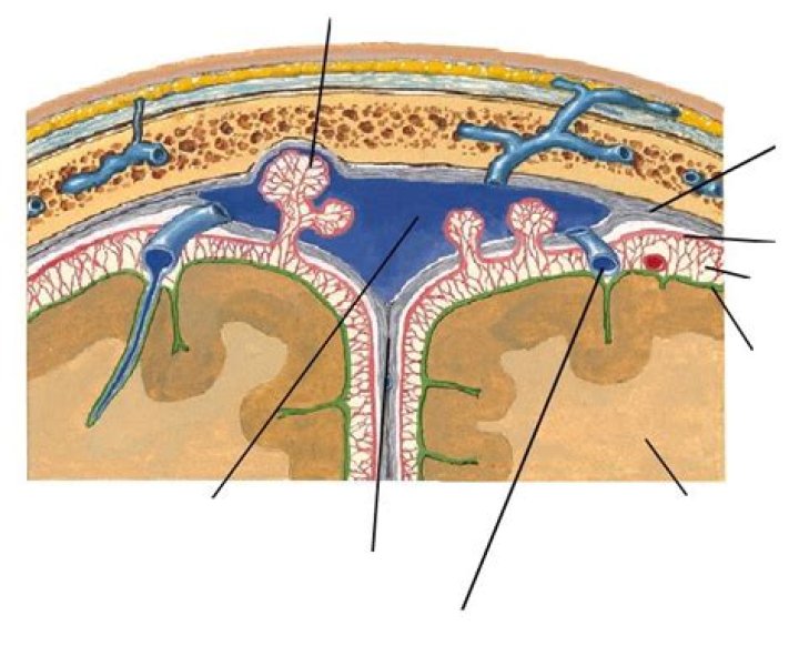
Arachnoid granulations (AGs) are tufts of arachnoid membrane invaginated into the dural sinuses through which cerebrospinal fluid (CSF) enters the venous system. The lesions are primarily located in the parasagittal region along the superior sagittal sinus[1], which is occasionally seen at the transverse sinus.
What happens if the arachnoid granulations are blocked?
Anytime there is a blockage in one of the channels of the brain or the arachnoid granulations, the plumbing system can get backed up. That backup can cause increased pressure in the brain because CSF is still produced in spite of the blockage. This condition is called hydrocephalus.
What causes arachnoid granulations?
When arachnoid villi become hypertrophied, possibly due to increased CSF pressure and volume, they transform into arachnoid granulations, typically greater than 2 mm [3]. These granulations are usually found in the superior sagittal sinus, but have been noted in other ectopic locations [4].Do arachnoid granulations enhance?
The key MRI features of giant arachnoid granulations are non-enhancing granules with central linear enhancement and surrounding enhancing flowing blood on contrast-enhanced MR venography3). Intrasinus thrombus may show contrast enhancement and occlude venous flow.
Can arachnoid granulations cause headaches?
Giant arachnoid granulations have been reported to be associated with headaches, which can be acute or chronic in presentation. In some cases, idiopathic intracranial hypertension, previously called pseudotumor cerebri, may occur.
Is arachnoid granulation normal?
They are focal, well-defined, and typically located within the lateral transverse sinuses adjacent to venous entrance sites. They should not be mistaken for sinus thrombosis or intrasinus tumor, but recognized as normal structures.
How many arachnoid granulations are there?
The average number of arachnoid granulations in the transverse sinuses is highest in the age group of 20 years (1.0 ± 1.4; H(6) = 14.48, p = 0.0247), while in other age groups, their average number in the transverse sinuses is equal to or less than 0.5.Do arachnoid cysts go away?
Most arachnoid cysts are stable and do not require treatment. They are four times more common in boys than in girls. Arachnoid cysts are diagnosed with a CT or MRI scan. Treatment, if necessary, involves draining the fluid through surgery or shunting.
How does CSF leave the brain?From the fourth ventricle, the CSF may exit through the foramen of Lushka laterally, or the foramen of Magendie medially to the subarachnoid space. Passing through the foramen of Magendie results in filling of the spinal subarachnoid space.
Article first time published onWhere do arachnoid granulations drain?
Arachnoid granulations are structures filled with cerebrospinal fluid (CSF) that extend into the venous sinuses through openings in the dura mater and allow the drainage of CSF from subarachnoid space into venous system.
Do arachnoid granulations allow for reabsorption of cerebrospinal fluid?
Absorption of CSF occurs across the arachnoid villi by a valve-like mechanism. Electron microscopic images of the arachnoid granulations show a series of channel-like structures [10]. The channels behave as one-way valves, allowing CSF to drain into the blood, but preventing blood from entering the CSF.
What is dura?
Dura: The outermost, toughest, and most fibrous of the three membranes (meninges) covering the brain and the spinal cord. Dura is short for dura mater (from the Latin for hard mother). … An accumulation of blood outside the dura is an epidural hematoma. Subdural means under the dura.
What does transverse sinus drain into?
The transverse sinuses are formed by the tentorium cerebelli and drain into the right and left sigmoid sinuses. … They drain from the confluence of sinuses (by the internal occipital protuberance) to the sigmoid sinuses, which ultimately connect to the internal jugular vein.
Where is the arachnoid mater?
The arachnoid mater, named for its spiderweb-like appearance, is a thin, transparent membrane surrounding the spinal cord like a loosely fitting sac. Continuous with the cerebral arachnoid above, it passes through the foramen magnum and descends caudally to the S2 vertebral level.
What is emissary vein?
The emissary veins are valveless vessels which connect the superficial veins of the scalp with deeper veins, e.g. diploic veins of the skull bones. From: Essential Clinical Anatomy of the Nervous System, 2015.
What is the arachnoid villi or arachnoid granulations of the?
Arachnoid granulations (also arachnoid villi, and pacchionian granulations or bodies) are small protrusions of the arachnoid mater (the thin second layer covering the brain) into the outer membrane of the dura mater (the thick outer layer).
What is the function of the arachnoid granulations quizlet?
Small protrusions of the arachnoid (the thin second layer covering the brain) through the dura mater (the thick outer layer). They protrude into the venous sinuses of the brain, and allow cerebrospinal fluid (CSF) to exit the sub-arachnoid space and enter the blood stream. Arachnoid granulations act as one-way valves.
What does the subdural space contain?
The classic view has been that a so-called subdural space is located between the arachnoid and dura and that subdural hematomas or hygromas are the result of blood or cerebrospinal fluid accumulating in this (preexisting) space.
What is dural sinus thrombosis?
A dural sinus thrombosis is the occlusion of a dural sinus by a blood clot (or thrombus). Because of this occlusion, blood flowing out of the brain is backed up, and the brain tissue becomes congested.
Where is the sagittal sinus?
The superior sagittal sinus (also known as the superior longitudinal sinus), within the human head, is an unpaired area along the attached margin of the falx cerebri. It allows blood to drain from the lateral aspects of anterior cerebral hemispheres to the confluence of sinuses.
What is lateral lacunae?
The lateral lacunae are small openings that communicate with irregularly shaped venous spaces (venous lacunae) in the dura mater near the sinus. There are usually three lacunae on either side of the superior sagittal sinus: a small frontal, a large parietal, and an occipital, intermediate in size between the other two.
What happens if an arachnoid cyst bursts?
With modern brain imaging studies, arachnoid cysts are often detected “incidentally”—during imaging tests performed for another reason. Although the cysts usually cause no harm, if they rupture (break open) or bleed, they can cause potentially serious problems requiring emergency treatment.
How serious is an arachnoid cyst?
Untreated, arachnoid cysts may cause permanent severe neurological damage when progressive expansion of the cyst(s) or bleeding into the cyst injures the brain or spinal cord. Symptoms usually resolve or improve with treatment.
Is arachnoid cyst life threatening?
Early detection and treatment of arachnoid cysts will help prevent symptoms from developing. If the cyst is allowed to grow, it may put pressure on the brain and spinal cord, leading to permanent neurological complications. Complications of untreated arachnoid cysts can be serious, even life threatening in some cases.
Why granulation is required?
Why is Granulation Necessary? The granulation process allows particles to stick together more firmly. It increases the particle size of the constituents used, which are mostly very fine powders. The greater the particle size of a constituent, greater will be its compressive or binding ability.
Which liquid is present in brain?
Cerebrospinal fluid is the liquid around your brain and spinal cord.
Does pia mater contain CSF?
Function. In conjunction with the other meningeal membranes, pia mater functions to cover and protect the central nervous system (CNS), to protect the blood vessels and enclose the venous sinuses near the CNS, to contain the cerebrospinal fluid (CSF) and to form partitions with the skull.
Does CSF regenerate?
After originating in the ventricles of the brain, it is probably filtered through the nervous-system membranes (ependyma). The CSF is continually produced, and all of it is replaced every six to eight hours.
What produces cerebrospinal fluid or CSF?
Cerebrospinal fluid (CSF) is a clear, colorless body fluid found within the tissue that surrounds the brain and spinal cord of all vertebrates. CSF is produced by specialised ependymal cells in the choroid plexus of the ventricles of the brain, and absorbed in the arachnoid granulations.
Does the subarachnoid space contain CSF?
The subarachnoid space consists of the cerebrospinal fluid (CSF), major blood vessels, and cisterns. The cisterns are enlarged pockets of CSF created due to the separation of the arachnoid mater from the pia mater based on the anatomy of the brain and spinal cord surface.