What is the uveal tract
By David Edwards
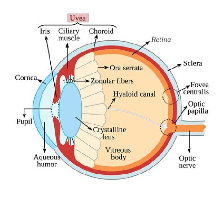
Also called uvea. … Enlarge. Anatomy of the eye, showing the outside and inside of the eye including the eyelid, pupil, sclera, iris, cornea, lens, ciliary body
What is the uveal tract of the eye?
The uveal tract is the middle layer of the eye, divided into the anterior uvea (iris, ciliary body) and posterior uvea (choroid). The uvea is sandwiched between an outer layer (sclera) and an inner layer (retina).
What is the function of the uvea?
The uvea is the middle layer of the eye. It lies beneath the white part of the eye (the sclera). It is made of the iris, ciliary body, and choroid. These structures control many eye functions, including adjusting to different levels of light or distances of objects.
Why is it called uveal tract?
The uveal tract, or simply uvea, is the pigmented middle membrane of the layers that make up the eye. The uveal tract is also called the vascular tunic of the eye because it is rich in its blood supply – i.e., vascular – and because it envelops the eye like a tunic would cover a body.Which part were inflammation of uveal tract include?
Uveitis is inflammation anywhere in the pigmented inside lining of the eye, known as the uvea or uveal tract. The uveal tract may become inflamed because of infection, injury, a bodywide autoimmune disorder (which causes the body to attack its own tissues), or for unknown reasons.
Is ciliary body transparent?
The ciliary body is a ring-shaped thickening of tissue inside the eye that divides the posterior chamber from the vitreous body. … The inner layer is transparent and covers the vitreous body, and is continuous from the neural tissue of the retina.
Why do you think the uveal coat is the middle coat?
The middle coat of the eye is called the uvea (from the Latin for “grape”) because the eye looks like a reddish-blue grape when the outer coat has been dissected away. The posterior part of the uvea, the choroid, is essentially a layer of blood vessels and connective tissue sandwiched between the sclera and the retina.
What is ora serrata in human eye?
The ora serrata is the peripheral termination of the retina and lies approximately 5 mm anterior to the equator of the eye. … The ora serrata is approximately 2 mm wide and is the site of transition from the complex, multilayered neural retina to the single, nonpigmented layer of ciliary epithelium.Is uvea same as choroid?
is that choroid is (anatomy) the vascular layer of the eye lying between the retina and the sclera while uvea is (anatomy) the middle of the three concentric layers that make up the eye; it is pigmented and vascular, and comprises the choroid, the ciliary body, and the iris.
Is the choroid the uvea?The uvea or vascular tunic of the eye consists of the choroid, ciliary body, and iris. The choroid lies between the sclera and RPE, and contains connective tissue, capillaries, and melanocytes. The choroid terminates anteriorly as the ciliary body.
Article first time published onWhat does Episcleritis look like?
Episcleritis often looks like pink eye, but it doesn’t cause discharge. It also may go away on its own. If your eye looks very red and feels painful, or your vision is blurry, seek immediate treatment.
What is granulomatous uveitis?
Granulomatous uveitis is an inflammation of the uveal tract characterized by the formation of granulomas due to infectious or non-infectious causes.
Does scleritis affect vision?
If it’s not treated, scleritis can lead to serious problems, like vision loss. It also can be linked to issues with your blood vessels (known as vascular disease).
Can Panuveitis be cured?
Panuveitis may be treated in several ways, including injections around the eye, oral medications, and eye drops. Corticosteroids are the treatment of choice for most types of uveitis, including panuveitis.
How long does it take to go blind from uveitis?
The mean duration of visual loss was 21 months. Of the 148 patients with pan-uveitis, 125 (84.45%) had reduced vision, with 66 (53%) having vision ⩽6/60.
What is inflammation What are the symptoms and signs of inflammation?
Symptoms of inflammation include: Redness. A swollen joint that may be warm to the touch. Joint pain.
What are human eyeballs made of?
It is made of water, jelly, and protein. The eyeball consists of these parts: Sclera.. The sclera is often referred to as the “whites of your eyes,” the tough white tissue that covers most of your eyeball.
What is the eyeball?
eyeball, spheroidal structure containing sense receptors for vision, found in all vertebrates and constructed much like a simple camera. … Much of the eyeball is filled with a transparent gel-like material, called the vitreous humour, that helps to maintain the spheroidal shape.
Which is the innermost coat of eyeball in rat?
The inner layer is the retina, which lines the back two-thirds of the eyeball.
What produces vitreous humor?
It is produced by the non-pigmented cells in the ciliary body. The vitreous humor fills the space (called vitreous chamber) between the lens and the retina of the eyeball. It is gelatinous near the edges and fluid-like near the center.
What Innervates ciliary body?
Innervation. The major innervation is provided by ciliary nerve branches (third cranial nerve-oculomotor), forming a rich parasympathetic plexus.
Is the ciliary body part of the choroid?
The ciliary body is the forward continuation of the choroid. It is a muscular ring, triangular in horizontal section, beginning at the region called the ora serrata and ending, in front, as the root of the iris. The surface is thrown into folds, called ciliary processes,…
What types of pathological processes of uvea do you know?
Common pathologic changes involving the uvea include inflammatory and neoplastic diseases. Inflammatory changes are clinically recognized as various forms of uveitis. Among the neoplasms, both primary and metastatic tumors are found in all parts of the uvea.
Where is uvea attached to sclera?
It has a rough outer surface which is attached to the sclera at the optic nerve and at the exit of the vortex veins. The smooth inner surface of the choroid is attached to the retinal pigmented epithelium (RPE). Choroid becomes continuous with pia and arachnoid at the optic nerve.
Where is uvea island located?
Uvea ʻUveaCountryFranceTerritoryWallis and FutunaIslandWallisCapitalMata-Utu
How is aqueous humor drained?
Aqueous humour drains out of the eye through the trabecular meshwork. The trabecular meshwork is a spongy mass of tiny canals located in the drainage angle. The drainage angle is located between the iris and the clear covering of the eye (cornea), where the iris meets the white outer covering (sclera) of the eye.
What tissue is the ora serrata made of?
Ora serrataTA98A15.2.04.006TA26780FMA58600Anatomical terminology
What is scleral spur?
The scleral spur is a fibrous ring that, on meridional section, appears as a wedge projecting from the inner aspect of the anterior sclera (Figs 3-1 and 3-2). The spur is attached anteriorly to the trabecular meshwork and posteriorly to the sclera and the longitudinal portion of the ciliary muscle.
Do humans have photoreceptor cells?
There are currently three known types of photoreceptor cells in mammalian eyes: rods, cones, and intrinsically photosensitive retinal ganglion cells. … In humans, there are three different types of cone cell, distinguished by their pattern of response to light of different wavelengths.
Is the sclera vascular?
The sclera is relatively inactive metabolically and has only a limited blood supply. Some blood vessels pass through the sclera to other tissues, but the sclera itself is considered avascular (lacking blood vessels).
What triggers episcleritis?
There is no apparent cause, but it can be associated with an underlying systemic inflammatory or rheumatologic condition such as rosacea, lupus or rheumatoid arthritis. Typical symptoms include generalized or local redness of the eyes that may be accompanied by mild soreness or discomfort but no visual problems.