What is unbalanced Avsd
By Victoria Simmons
Unbalanced atrioventricular septal defect (uAVSD) is a challenging lesion with suboptimal outcomes in the current era. Severe forms of uAVSD mandate univentricular repair with well-documented outcomes. Determining the feasibility of biventricular repair (BVR) in patients with moderate forms of uAVSD is difficult.
What is an unbalanced AV canal defect?
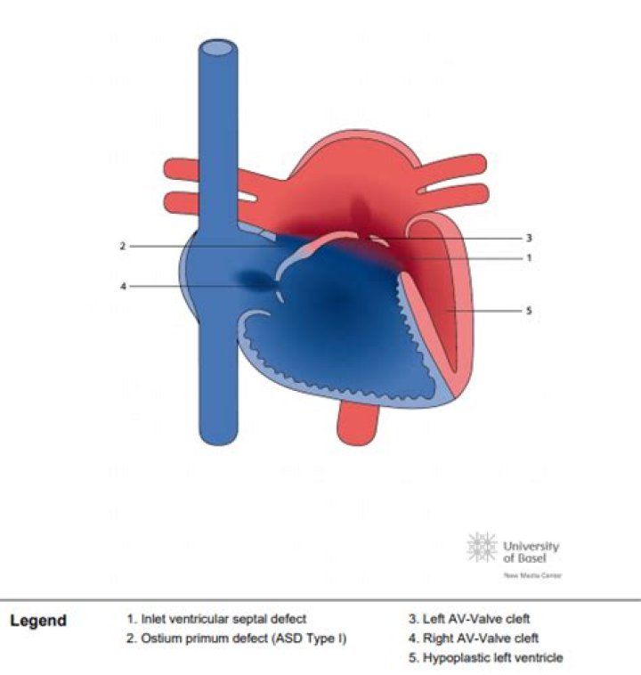
Although a considerable spectrum of ventricular dominance occurs, the term unbalanced AV septal defect generally implies hypoplasia of one ventricle and its associated outflow tract with essentially single-ventricle physiology. RV-dominant AV septal defects occur more commonly than LV-dominant AV septal defects.
What is the difference between Avsd and ASD?
Atrioventricular septal defect (AVSD), also referred to as endocardial cushion defects, consists of three defects in the heart: An atrial septal defect (ASD), a hole in the “wall” (septum) of the heart that separates the two upper chambers (the atria).
How many types of Avsd are there?
There are two types of AVSD: Partial AVSD: A hole exists in the wall between the heart’s upper chambers, and the valve between the left chambers does not close completely. Complete AVSD: A large hole exists in the center of the heart where the walls between the upper and lower chambers meet.Can you have Avsd without Down syndrome?
In patients with AVSD without Down syndrome, about 20% have other genetic disorders such as Holt-Oram or Noonan syndrome.
What is an AVC heart?
Atrioventricular canal defect is a type of congenital heart defect. A person born with atrioventricular canal defect has a hole in the wall separating the heart’s chambers and problems with the heart valves. The condition may be partial, involving only the two upper chambers, or complete, involving all four chambers.
How do you treat atrioventricular canal defect?
Surgery is needed to repair a complete or partial atrioventricular canal defect. More than one surgery may be needed. Surgery to correct atrioventricular canal defect involves using one or two patches to close the hole in the heart wall.
Does AVSD cause cyanosis?
Some babies with AVSD look a little blue in the lips and/or under their fingernails, especially when they cry. This is called cyanosis and occurs when blood on the right side of heart flows to the left side of the heart (and out to the body) through one of the holes.Is AVSD cyanotic or Acyanotic?
Infants with complete atrioventricular septal defect often have a bluish discoloration of the skin and mucous membranes (cyanosis) due to insufficient oxygen supply to these tissues.
Can ventricular septal defect be prevented?In most cases, you can’t do anything to prevent having a baby with a ventricular septal defect. However, it’s important to do everything possible to have a healthy pregnancy.
Article first time published onDoes Avsd require surgery?
All AVSDs, both partial and complete types, usually require surgery. During surgery, any holes in the chambers are closed using patches. If the mitral valve does not close completely, it is repaired or replaced.
What is the difference between VSD and Avsd?
In AV septal defect: There is a hole in the wall between the right and left atria (atrial septal defect, ASD). There is a hole in the wall between the right and left ventricles (ventricular septal defect, VSD).
Is Avsd the same as ASD and VSD?
A partial AVSD is defined as a common atrioventricular junction with two separate orifices, a left AVV with three leaflets (cleft) and a primum ASD (Jacobs et al., 2000a). A complete AVSD is defined as a primum ASD, inlet VSD, and a large common valve that spans the defects (Jacobs et al., 2000a).
Is AVSD hereditary?
The genetic etiology of atrioventricular septal defect (AVSD) is unknown in 40% cases. Conventional sequencing and arrays have identified the etiology in only a minority of nonsyndromic individuals with AVSD.
Do all babies with AVSD have Down syndrome?
ATRIOVENTRICULAR SEPTAL DEFECT (AVSD) AVSD is the most frequently diagnosed congenital heart condition in children with Down syndrome. Various studies place the incidence rate between 30 and 47 percent of CHDs in children with Down syndrome, according to the book Advances in Research on Down Syndrome.
What does truncus arteriosus mean?
Truncus arteriosus is a birth defect of the heart. It occurs when the blood vessel coming out of the heart in the developing baby fails to separate completely during development, leaving a connection between the aorta and pulmonary artery.
What is Cuspid valve?
Valves of the Heart The heart has two types of valves that keep the blood flowing in the correct direction. The valves between the atria and ventricles are called atrioventricular valves (also called cuspid valves), while those at the bases of the large vessels leaving the ventricles are called semilunar valves.
What is Rastelli type A?
Rastelli Type A (Figures 8 and 9, Videos 20 and 21). This is the most common type of complete CAVC (75%). The superior bridging leaflet is completely divided from the free edge to the annulus at the level of the ventricular septum.
Why is Avsd common in Down syndrome?
In Down syndrome, complete AVSD is often seen. The increased adhesiveness of trisomy 21 cells might keep the embryonal endocardial cushion from fusing, thereby causing persistent AVSD.
Why ASD is Acyanotic?
Acyanotic heart defectVentricular septumSpecialtyCardiology
Why is Acyanotic left to right?
With physiologic declines in pulmonary vascular resistance, compensatory in utero right ventricular hypertrophy regresses, resulting in a more compliant right ventricle and atrium. This allows a progressive left-to-right increase in ASD shunt volume that is further pronounced with larger defect size.
Can heart muscles repair?
But the heart does have some ability to make new muscle and possibly repair itself. The rate of regeneration is so slow, though, that it can’t fix the kind of damage caused by a heart attack. That’s why the rapid healing that follows a heart attack creates scar tissue in place of working muscle tissue.
Why does VSD cause shortness of breath?
Because the heart and lungs have to work harder, a baby with a ventricular septal defect will become short of breath, particularly with the exertion of feeding (which is the most exercise a baby does). This could lead to poor feeding and eventually to poor weight gain and growth.
Can a closed hole in your heart reopen?
There are no known medications that can repair the hole. If a child is diagnosed with an atrial septal defect, the health care provider may want to monitor it for a while to see if the hole closes on its own. During this period of time, the health care provider might treat symptoms with medicine.
Can you live a normal life with a hole in your heart?
It is very possible to live with a hole in your heart, without ever realising that it’s there. A patent foramen ovale, also known as a PFO, is a hole between the left and right atria (upper chambers) of the heart that we all have when we are in the womb, but this should close shortly after we’re born.
How long does an AVSD repair take?
The repair will take about 2 hours. The healthcare provider puts a small, flexible tube (catheter) into several blood vessels in the groin. One of the catheters will have a small device inside it. The provider threads the catheter through the blood vessel all the way to the ventricular septum.
What does AVSD murmur sound like?
A large AVSD with substantial left-to-right shunting creates a mid-diastolic rumbling murmur, audible along the lower left sternal border. This often occurs in association with a prominent third heart sound (S3) in that location.
What is transitional AVSD?
Transitional AV Canal (aka intermediate AV canal) A transitional AVSD has two separate AV valve annuli. In addition to a primum ASD and a cleft mitral valve, there is often a small inlet VSD that is often restricted or obliterated by dense chordal attachments of the AV valves to the crest of the ventricular septum.
Is ASD life threatening?
Severe cases of atrial septal defects may lead to life-threatening complications such as chest pain, irregular heartbeats (arrhythmias), abnormal enlargement of the heart, a “fluttering” of the heart (atrial fibrillation), and/or heart failure.