What is villous tissue
By Christopher Green
villus, plural villi, in anatomy any of the small, slender, vascular projections that increase the surface area of a membrane. Important villous membranes include the placenta and the mucous-membrane coating of the small intestine.
What is the villous?
Definition of villous 1 : covered or furnished with villi a villous adenoma. 2 : having soft long hairs leaves villous underneath — compare pubescent.
What is villi pregnancy?
During pregnancy, the placenta provides oxygen and nutrients to the growing baby and removes waste products from the baby’s blood. The chorionic villi are wispy projections of placental tissue that share the baby’s genetic makeup. The test can be done as early as 10 weeks of pregnancy.
Are villi tissues?
The bulk of the villi consist of connective tissues that contain blood vessels. Most of the cells in the connective tissue core of the villi are fibroblasts. Macrophages known as Hofbauer cells are also present.What is villous formation?
Villi formation occurs by the invagination of the mesenchymal cells that underlie the luminal epithelia, a process that follows the anterior–posterior wave of epithelial reorganization. These mesenchymal cells condense under the basal lamina and grow in fingerlike projections toward the lumen giving rise to the villi.
What are villi and microvilli?
Microvilli is a part of a cell. Its function is to augment the surface area of the cell. The main function of microvilli includes secretion, absorption, and cellular sticking or adhesion. Villi or intestinal villi, on the other hand, are finger-like projections that are found in the intestinal wall.
How do villi work?
Villi are specialized for absorption in the small intestine as they have a thin wall, one cell thick, which enables a shorter diffusion path. They have a large surface area so there will be more efficient absorption of fatty acids and glycerol into the blood stream.
Where is the Rugae?
The rugae are folds in the stomach lining.What is villous chorion?
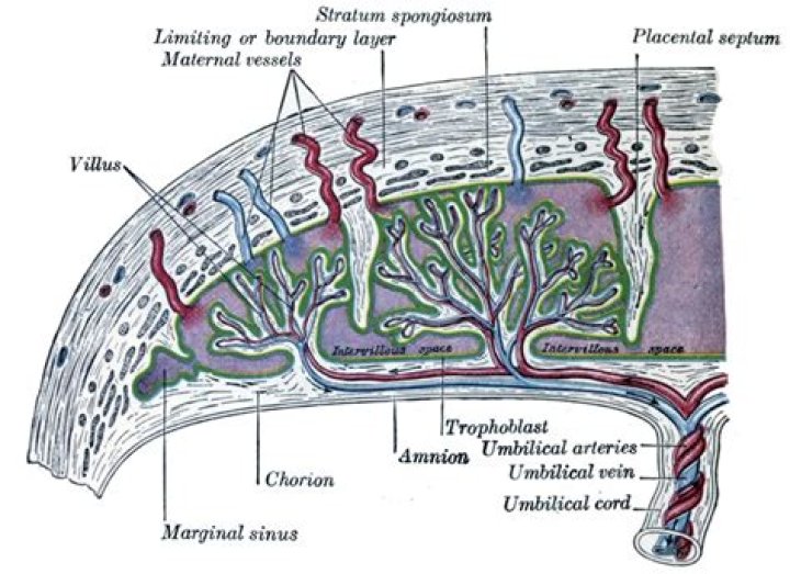
The fetal portion of the placenta is known as the villous chorion. The maternal portion is known as the decidua basalis. The two portions are held together by anchoring villi that are anchored to the decidua basalis by the cytotrophoblastic shell.
Where is villi found?They are most prevalent at the beginning of the small intestine and diminish in number toward the end of the tract. They range in length from about 0.5 to 1 mm (about 0.02 to 0.04 inch). The large number of villi give the internal intestinal wall a velvety appearance.
Article first time published onWhat is Accreta?
Placenta accreta is a serious pregnancy condition that occurs when the placenta grows too deeply into the uterine wall. Typically, the placenta detaches from the uterine wall after childbirth. With placenta accreta, part or all of the placenta remains attached. This can cause severe blood loss after delivery.
What is missed abortion?
A missed abortion is a nonviable intrauterine pregnancy that has been retained within the uterus without spontaneous abortion. Typically, no symptoms exist besides amenorrhea, and the patient finds out that the pregnancy stopped developing earlier when a fetal heartbeat is not observed or heard at the appropriate time.
What is umbilical cord?
The umbilical cord is a flexible, tube-like structure that, during pregnancy, connects the fetus to the mother. The umbilical cord is the baby’s lifeline to the mother. It transports nutrients to the baby and also carries away the baby’s waste products. It is made up of three blood vessels – two arteries and one vein.
What is placental plate?
The fetal part of the placenta is made up of the chorionic plate with its placental villi, the cytotrophoblast layer and the intervillous spaces. The chorionic plate (great part of the placenta on the fetal side) consists of the amnion, the extra-embryonic mesenchyma, the cytotrophoblast and the syncytiotrophoblast.
What is the difference between chorionic villi and placental villi?
chorionic villi: These sprout from the chorion in order to give a maximum area of contact with the maternal blood. placenta: A vascular organ present only in the female during gestation. It supplies food and oxygen from the mother to the fetus, and passes back waste.
What is the function of placental villi?
villi in the placenta Chorionic villi make up a significant portion of the placenta and serve primarily to increase the surface area by which products from the maternal blood are made available to the fetus.
What is bile juice?
Bile juice is a digestive fluid produced by the liver. It is stored and concentrated in the gallbladder. Its main function is to convert fats in food into fatty acids, which are absorbed in the gut.
Where is the ileal?
The last part of the small intestine. It connects to the cecum (first part of the large intestine). The ileum helps to further digest food coming from the stomach and other parts of the small intestine.
Which is not digest by human?
Cellulose is a fibre which is not digestible by the human digestive system.
What is brush border?
The brush border is a complex and highly plastic organelle required for intestinal homeostasis and is specialized for absorption of nutrients. Thousands of tightly packed microvilli form the brush border together with the area they are located on, the so-called terminal web.
What are Paneth cells?
Paneth cells are highly specialized secretory epithelial cells located in the small intestinal crypts of Lieberkühn. The dense granules produced by Paneth cells contain an abundance of antimicrobial peptides and immunomodulating proteins that function to regulate the composition of the intestinal flora.
What is the function of goblet cells?
Goblet cells reside throughout the length of the small and large intestine and are responsible for the production and maintenance of the protective mucus blanket by synthesizing and secreting high-molecular-weight glycoproteins known as mucins.
What is the difference between chorion and amnion?
The amnion is found on the innermost part of the placenta. It lines the amniotic cavity and holds the amniotic fluid and the developing embryo. … The chorion, on the other hand, is the outer membrane that surrounds the amnion, the embryo, and other membranes and entities in the womb.
How serious is vasa previa?
Vasa previa doesn’t pose any physical health risks to the mother, but the risks to the baby can be significant and can ultimately result in the loss of their life. More than half of all cases of vasa previa that aren’t detected in pregnancy result in stillbirth.
Is chorion the same as placenta?
The main difference between chorion and placenta is that chorion is the outermost fetal membrane, covering the embryo of mammals, reptiles, and birds whereas placenta is the temporary organ that connects the developing fetus to the uterine wall through umbilical cord in mammals.
What is pyloric sphincter?
The pyloric sphincter muscle is responsible for controlling how partially digested food, called chyme, moves from your stomach and into your intestines in a timely manner. This process, known as gastric emptying, should happen at an optimal rate to ensure good digestion.
What is gastric fold?
The gastric folds (or gastric rugae) are coiled sections of tissue that exist in the mucosal and submucosal layers of the stomach. They provide elasticity by allowing the stomach to expand when a bolus enters it.
What is mucosal fold?
A mucosal fold refers to a fold in any mucous membrane in the body. This may refer to: Gastric fold of the gastric mucosa. Transverse folds of rectum in the anal canal. Circular folds in the small intestine.
Which organ removes water from undigested?
The large intestine’s main job is to remove water from the undigested matter and form solid waste (poop) to be excreted. The large intestine has three parts: The cecum (pronounced: SEE-kum) is the beginning of the large intestine.
Where is enzyme produced?
Enzymes are produced naturally in the body. For example, enzymes are required for proper digestive system function. Digestive enzymes are mostly produced in the pancreas, stomach, and small intestine.
Where does the blood leaving the ileum flow to?
Nutrient-rich blood flows into the liver from the intestines through the hepatic portal vein.