What nerve Innervates the ankle
By David Edwards
Tibial nerve: Rami branch out to the ankle joint in the distal third of the lower leg. In addition, branches extend from the medial plantar nerve to the ankle joint.
What nerve affects the ankle?
The tibial nerve runs through the tarsal tunnel, which is a narrow passageway inside your ankle that is bound by bone and soft tissue. Damage of the tibial nerve typically occurs when the nerve is compressed as a result of consistent pressure.
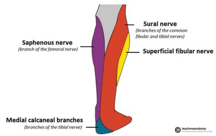
What nerve innervates the lateral ankle?
Sural nerveFromMedial sural cutaneous nerve, communicating branch with the common fibular nerve (S1, S2)InnervatesSupplies sensation to the skin of the lateral foot and lateral lower ankle.IdentifiersLatinnervus suralis
What are symptoms of peroneal nerve damage?
- Decreased sensation, numbness, or tingling in the top of the foot or the outer part of the upper or lower leg.
- Foot that drops (unable to hold the foot up)
- “Slapping” gait (walking pattern in which each step makes a slapping noise)
What does nerve damage in the ankle feel like?
Damage to the peroneal nerve can cause pain, tingling or numbness at the top of the foot. It may become difficult to raise your toes, your toes or ankle may feel weak, or your foot may feel like it is dropping when walking. In severe cases, you may be completely unable to lift your toes or foot or turn your ankle.
What is Baxter's nerve?
The Baxter’s nerve, also known the first branch of the lateral plantar nerve, is a small nerve (under 1mm in diameter) running along the inside of the heel. It is an uncommon cause of heel pain, but one that should be considered when pain is not improving, particularly if you have been diagnosed with plantarfasciitis.
What is neuroma?
Neuroma. A neuroma is a disorganized growth of nerve cells at the site of a nerve injury. A neuroma occurs after a nerve is partially or completely disrupted by an injury — either due to a cut, a crush, or an excessive stretch.
What causes peroneal nerve inflammation?
Common causes of damage to the peroneal nerve include the following: Trauma or injury to the knee. Fracture of the fibula (a bone of the lower leg) Use of a tight plaster cast (or other long-term constriction) of the lower leg.How do you test for peroneal nerve damage?
To test for the motor involvement of the superficial peroneal nerve and deep peroneal nerve, one must assess foot eversion (SPN) and foot/toe dorsiflexion (DPN). A finding of weakness of both foot eversion as well as foot/toe dorsiflexion suggests a lesion involving the common peroneal nerve.
What does saphenous nerve innervate?Anatomy. The saphenous nerve is the largest branch of the femoral nerve and innervates the medial portion of the lower leg and the foot. The nerve travels with the femoral artery through Hunter’s canal, and at the medial condyle of the distal femur it supplies the medial portion of the knee and the lower leg.
Article first time published onWhat is superficial peroneal nerve?
The superficial peroneal nerve (superficial fibular nerve) is a mixed nerve that carries sensory information from the anterolateral aspect of the leg and the greater part of the dorsum of the foot (except for the first web space).
Where is the peroneal nerve?
The peroneal nerve is on the outside of the fibula just below the knee. Pressure to the peroneal nerve, as you might experience if you sit with your legs crossed for too long, can trigger temporary foot drop.
How do you fix nerve damage in your foot?
- Braces or splints. These devices keep the affected limb, fingers, hand or foot in the proper position to improve muscle function.
- Electrical stimulator. …
- Physical therapy. …
- Exercise.
Can a pinched nerve be detected with an MRI?
MRI scans which show soft tissues, such as nerves and discs, are generally preferred over CT scans which show bony elements. Advanced imaging can show exactly which nerve or nerves are being pinched and what is causing the nerve to be pinched.
What causes sharp pain in ankle bone?
The most common causes include injury, arthritis and normal wear and tear. Depending on the cause, you may feel pain or stiffness anywhere around the ankle. Your ankle may also swell, and you may not be able to put any weight on it. Usually, ankle pain gets better with rest, ice and over-the-counter pain medications.
What is Neutris?
Neuritis is a medical term meaning inflammation in the peripheral nerves (nerves outside the central nervous system are called peripheral).
What is distal stump?
The distal stump refers to the end of the injured neuron that is still attached to the end of the axon; it is the part of the neuron that will degenerate, but the stump remains capable of regenerating its axons.
What is tarsal syndrome?
What Is Tarsal Tunnel Syndrome? Tarsal tunnel syndrome is a compression, or squeezing, on the posterior tibial nerve that produces symptoms anywhere along the path of the nerve running from the inside of the ankle into the foot. Tarsal tunnel syndrome is similar to carpal tunnel syndrome, which occurs in the wrist.
What is plantar nerve entrapment?
Medial and lateral plantar nerve entrapment is compression of nerve branches at the inner heel (the medial or lateral branches of the plantar nerve) that causes pain. The nerve branches are pinched (compressed) between bone, ligaments, and other connective tissues, causing pain.
What does Baxter's nerve entrapment feel like?
Patients with Baxter’s neuritis often experience numbness along the inside of the heel (medial aspect). This is because the nerve being entrapped enters the foot behind the inside ankle bone and dives deep into the foot beneath the muscle that fills the arch (abductor hallucis muscle).
Where does Baxter's nerve entrapment hurt?
In Baxter’s Nerve Entrapment distinct tenderness is felt at the origin of the abductor hallicus muscle (a small muscle along the inside of the foot), whereas the most intense site of pain in Plantar Fasciitis is more commonly towards the bottom of the heel.
Where does common peroneal nerve split?
It divides at the knee into two terminal branches: the superficial peroneal nerve and deep peroneal nerve, which innervate the muscles of the lateral and anterior compartments of the leg respectively.
What causes compression of peroneal nerve?
Peroneal neuropathies are classically associated with external compression at the level of the fibular head. The most common etiology is habitual leg crossing (which compresses this area). Prolonged positioning with pressure at this area (e.g. sitting on an airplane or positioning during surgery) are other causes.
How long does it take the peroneal nerve to heal?
The recovery time after a common peroneal nerve decompression at the knee is usually 3-4 months. For the first 6 weeks, we do not want to encourage the knee to form a lot of scar tissue around the area of the decompression, so we have patients on crutches.
What muscles does common peroneal nerve innervate?
- Tibialis anterior.
- Extensor digitorum longus.
- Peroneus tertius.
- Extensor hallucis longus (propius)
- Extensor digitorum brevis.
- Extensor hallucis brevis.
How do you fix peroneal nerve damage?
Nonsurgical treatments, including orthotics, braces or foot splints that fit inside the person’s shoe, can bring relief. Physical therapy and gait retraining can help the person improve their mobility. Some injuries may require peripheral nerve surgery, including one or more of these procedures: Decompression surgery.
What does the femoral nerve innervate?
In the proximal thigh, the femoral nerve divides into sensory branches, which innervate the upper and anterior thigh, and muscular branches, which innervate the quadriceps. One of the major branches is the lateral femoral cutaneous nerve.
What does the common fibular nerve innervate?
The common fibular nerve innervates the short head of the biceps femoris muscle (part of the hamstring muscles, which flex at the knee).
What does the tibial nerve innervate?
In addition to the two heads of the gastrocnemius muscle, the tibial nerve innervates the plantaris, soleus, popliteus, posterior tibialis, flexor digitorum longus, and flexor hallucis longus muscles (Fig. … The medial and lateral plantar nerves carry motor and sensory fibers.
What nerve root Innervates dorsum of foot?
The superficial peroneal nerve, sometimes called the superficial fibular nerve, innervates the peroneus longus and peroneus brevis muscles and the skin over the antero-lateral aspect of the leg along with the greater part of the dorsum of the foot (with the exception of the first web space, which is innervated by the …
Where does superficial fibular nerve originate?
The superficial branch of the common peroneal nerve, also known as the superficial fibular (peroneal) nerve, arises at the bifurcation of the common peroneal nerve, between the fibula and the proximal part of fibularis longus muscle. This nerve then descends deep to the fibularis longus muscle.