What part of the ECG represents ventricular repolarization
By James Craig
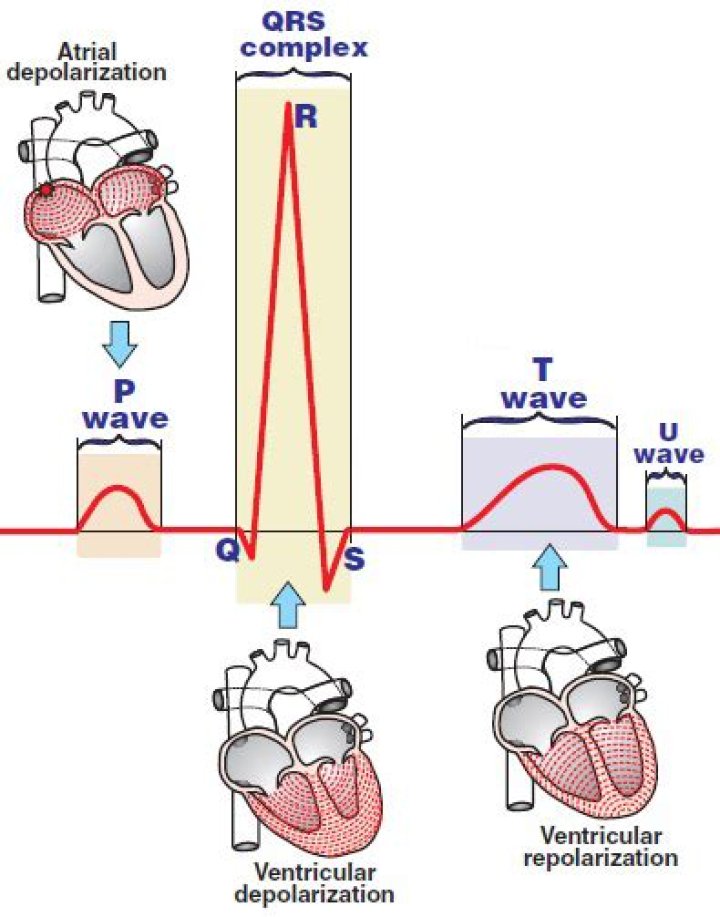
The T wave represents ventricular repolarization. Generally, the T wave exhibits a positive deflection. The reason for this is that the last cells to depolarize in the ventricles are the first to repolarize.
What part of the ECG represents ventricular repolarization quizlet?
The T wave represents ventricular repolarization. The P wave represents atrial depolarization.
What is repolarization on ECG?
Early repolarization pattern (ERP) is a common ECG variant, characterized by J point elevation manifested either as terminal QRS slurring (the transition from the QRS segment to the ST segment) or notching (a positive deflection inscribed on terminal QRS complex) associated with concave upward ST-segment elevation and …
Does the T wave represent ventricular repolarization?
The T wave on the ECG (T-ECG) represents repolarization of the ventricular myocardium. Its morphology and duration are commonly used to diagnose pathology and assess risk of life-threatening ventricular arrhythmias.What is ventricular repolarization?
Ventricular repolarization is a complex electrical phenomenon which represents a crucial stage in electrical cardiac activity. It is expressed on the surface electrocardiogram by the interval between the start of the QRS complex and the end of the T wave or U wave (QT).
What does each part of the ECG represent?
There are three main components to an ECG: the P wave, which represents the depolarization of the atria; the QRS complex, which represents the depolarization of the ventricles; and the T wave, which represents the repolarization of the ventricles.
What part of the ECG reflects the depolarization of the ventricles and repolarization of the atria?
Atrial and ventricular depolarization and repolarization are represented on the ECG as a series of waves: the P wave followed by the QRS complex and the T wave. The first deflection is the P wave associated with right and left atrial depolarization. Wave of atrial repolarization is invisible because of low amplitude.
What does QRS mean in ECG?
The QRS complex represents the electrical impulse as it spreads through the ventricles and indicates ventricular depolarization. As with the P wave, the QRS complex starts just before ventricular contraction.What does the QRS represent on an ECG?
The QRS wave is produced by the atrioventricular node (AV). The P wave in an ECG complex indicates atrial depolarization. The QRS is responsible for ventricular depolarization and the T wave is ventricular repolarization.
Where does repolarization of the heart occur?circulatory system. This repolarization process occurs in the muscle of the ventricles about 0.25 second after depolarization. There are, therefore, both depolarization and repolarization waves represented in the electrocardiogram.
Article first time published onWhere is the J point on ECG?
Introduction. The J-point on the electrocardiographic waveform is historically defined as the junction between the end of the QRS complex and the beginning of the ST-segment.
Where does atrial repolarization occur on an ECG trace?
There is no distinctly visible wave representing atrial repolarization in the ECG because it occurs during ventricular depolarization. Because the wave of atrial repolarization is relatively small in amplitude (i.e., has low voltage), it is masked by the much larger ventricular-generated QRS complex.
Which part of the QRS complex represents the repolarization of the atria?
The QRS complex represents ventricular depolarization (and atrial repolarization) or the journey through the electrical impulse from the Av-Node through the Purkinje network. (brought on by depolarization) and the beginning of electrical recovery (repolarization) is indicated by the S-T Segment.
Why is atrial repolarization not observed in the ECG?
There is no distinctly visible wave representing atrial repolarization in the ECG because it occurs during ventricular depolarization. Because the wave of atrial repolarization is relatively small in amplitude (i.e., has low voltage), it is masked by the much larger ventricular-generated QRS complex.
Which part of the waveform represents atrioventricular conduction?
The PR segment represents the electrical conduction through the atria and the delay of the electrical impulse in the atrioventricular node. After the signal leaves the AV node it travels along a pathway called the bundle of His (3) and into the right and left bundle branches (4, 5).
Which rhythm originates from the ventricle?
Ventricular arrhythmias are abnormal heart rhythms that originate in the bottom chambers of the heart called the ventricles.
Which of the following is associated with Osborn or J waves on the ECG?
Osborn wave (J wave). These waves occur due to hypothermia, hypercalcemia, early repolarization and Brugada syndrome.
Which of the following ECG waveforms represents ventricular depolarization?
The QRS complex represents ventricular depolarization.
Which valves are open during ventricular diastole?
The mitral and tricuspid valves, also known as the atrioventricular, or AV valves, open during ventricular diastole to permit filling.
Why is atrial repolarization not illustrated on an ECG quizlet?
Why is atrial repolarization not observed in the ECG? Atrial repolarization occurs at the same time as ventricular depolarization, the QRS complex hides atrial repolarization.
How many valves are closed in ventricular systole?
There are four heart valves that allow the flow from the atria to the ventricles and from the ventricles through to the arteries but do not allow the blood to flow back into either of the chambers.