
What is a circumscribed hypoechoic mass
By Olivia Hensley
A hypoechoic mass is tissue in the body that’s more dense or solid than usual. This term is used to describe what is seen on an ultrasound scan. Ultrasound uses sound waves that are absorbed by or bounce off of tissues, organs, and muscles.
Can a circumscribed mass be cancer?
Circumscribed oval and round masses are usually benign. An irregular shape suggests a greater likelihood of malignancy.
What is a well circumscribed mass?
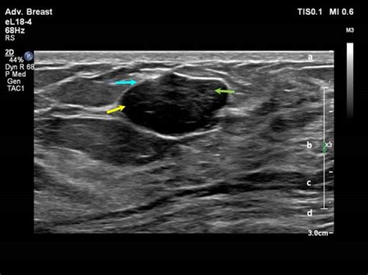
Circumscribed masses. A circumscribed mass in mammography is a mass where the contour is clearly defined along at least 75% of its surface. The remaining 25% may, at most, be masked by the adjacent gland. Circumscribed masses first indicate benign lesions.
Is hypoechoic mass cancerous?
Solid masses are hypoechoic and can be cancerous. Cysts filled with air or fluid are usually hyperechoic and are rarely cancerous. Abnormal tissue also looks different from healthy tissue on a sonogram. Your doctor will usually do further testing if an ultrasound shows a solid mass or what looks like abnormal tissue.Can a hypoechoic nodule be benign?
Spongiform nodules, purely or predominantly cystic nodules, nodules with well-defined hypoechoic halo and echogenic as well as isoechoic nodules are usually benign. None of the US characteristics have 100% accuracy in detecting or excluding malignancy.
What does a mass with circumscribed margins mean?
As defined in the US lexicon (1), circumscribed margins are well defined or sharp, with an abrupt transition between the lesion and the surrounding tissue. Noncircumscribed margins encompass the remaining margin descriptors, including microlobulated, indistinct, angular, and spiculated.
Is hypoechoic mass in breast cancer?
Breast cancer is the second leading cause of cancer-related death in women. Regular breast exams and screening are important. However, most growths found in the breast are benign. Most benign and malignant masses in the breast are hypoechoic.
What is the normal size of hypoechoic lesion?
The lesions measured from 6 to 20mm (mean 13.1 mm).What percentage of hypoechoic masses are malignant?
In addition increase in vascularity in the hypoechoic mass predicts malignancy about 82% of the time. The ultrasound image below shows an irregular vascularized retroareolar mass, with calcifications. This is very likely to be infiltrating ductal carcinoma and your doctor will recommend a biopsy straight away.
What causes hypoechoic mass in breast?In this review article, we classify benign breast lesions that show irregular hypoechoic masses on US into 4 groups: iatrogenic or trauma-related breast lesions (foreign body reaction, fat necrosis, fibrotic scar), inflammations (abscess, idiopathic granulomatous lobular mastitis [IGLM], diabetic mastopathy [DMP]), …
Article first time published onCan a doctor tell if a tumor is cancerous by looking at it?
Cancer is nearly always diagnosed by an expert who has looked at cell or tissue samples under a microscope. In some cases, tests done on the cells’ proteins, DNA, and RNA can help tell doctors if there’s cancer. These test results are very important when choosing the best treatment options.
Can a solid mass in the breast be benign?
Fortunately, a majority of breast lumps are benign, meaning they’re not cancerous. Both women and men can develop benign (noncancerous) breast lumps. This condition is known as benign breast disease.
What is a stable mass?
Cancer doctors use the term stable disease to describe a tumor that is neither growing nor shrinking. Specifically, it means that there was neither an increase in size of more than 20% nor a decrease in size of more than 30% since the initial baseline measurement.
How often are hypoechoic nodules cancerous?
While most thyroid nodules are non-cancerous (Benign), ~5% are cancerous. Thyroid Ultrasound: a common imaging test used to evaluate the structure of the thyroid gland.
How are hypoechoic nodules treated?
These type of nodules are usually solid rather than a fluid-filled lesion. If a doctor suspects that a thyroid nodule may be cancerous, they will recommend additional testing, such as blood tests and biopsies. The main treatment for cancerous nodules is surgical removal of part or all of the thyroid gland.
What is hypoechoic nodules?
A hypoechoic nodule is an area of swelling or abnormal cell growth on the thyroid. The term “hypoechoic” refers to the way the nodule appears on an ultrasound: dark. When a nodule appears hypoechoic rather than anechoic, radiologists know it’s likely solid and not liquid-filled.
Are breast cysts hypoechoic?
The majority of breast lesions detected by ultrasound are hypoechoic. According to the BI-RADS lexicon [1], a hyperechoic lesion is defined by an echogenicity greater than that of subcutaneous fat or equal to that of fibroglandular parenchyma.
What does a cancerous breast lump look like on an ultrasound?
On ultrasound, a breast cancer tumor is often seen as hypoechoic. It has irregular borders, and may appear spiculated. Other ultrasound findings that suggest breast cancer include: Nonparallel orientation (not parallel to the skin)
Why is a biopsy done on the breast?
Breast biopsies may be done: To check a lump or mass that can be felt (is palpable) in the breast. To check a problem seen on a mammogram, such as small calcium deposits in breast tissue (microcalcifications) or a fluid-filled mass (cyst) To evaluate nipple problems, such as a bloody discharge from the nipple.
Are dense breasts common?
How common are dense breasts? Nearly half of all women age 40 and older who get mammograms are found to have dense breasts. Breast density is often inherited, but other factors can influence it. Factors associated with lower breast density include increasing age, having children, and using tamoxifen.
What is Birads score?
The BI-RADS score is an acronym for the Breast Imaging Reporting and Database System score. It’s a scoring system radiologists use to describe mammogram results. A mammogram is an X-ray imaging test that examines breast health. It’s the most efficient tool to help detect breast cancer, especially at its earliest stage.
What does Birads 4 mean?
BI-RADS category 4 means there is a suspicious abnormality on your breast imaging studies and a biopsy should be considered as a next step. Remember the only way to actually diagnose breast cancer is to obtain a tissue sample for evaluation by a pathologist, a doctor specializing in looking at tissue samples.
What is Birads III?
BI-RADS 3 is an evolving assessment category. When used properly, it reduces the number of benign biopsies while allowing the breast imager to maintain a high sensitivity for the detection of early stage breast cancer.
Is a lipoma hypoechoic?
These soft-tissue masses are lower in reflectivity than muscle but more reflective than adjacent subcutaneous fat. In reality, while the majority of lipomas are hyperechoic, a significant proportion of them can also be hypoechoic or isoechoic (5).
What is the difference between hypoechoic and hyperechoic?
Hypoechoic: Gives off fewer echoes; they are darker than surrounding structures. Examples include lymph nodes and tumors. Hyperechoic: Increased density of sound waves compared to surrounding structures. Examples include bone and fat calcifications.
What does hyperechoic lesion mean?
According to the BI-RADS lexicon [1], a hyperechoic lesion is defined by an echogenicity greater than that of subcutaneous fat or equal to that of fibroglandular parenchyma. Only 1–6% of breast masses are hyperechoic and the great majority of them are benign.
What does it mean if a mass is echogenic?
An echogenic mass was defined as a well-circumscribed mass, often with a lobulated appearance and calcifications, without any fluid components.
Can a suspicious breast lump be benign?
If you find a breast lump or other change in your breast, you might worry about breast cancer. That’s understandable. But breast lumps are common, and most often they’re noncancerous (benign), particularly in younger women.
Are fibroadenomas hypoechoic?
Ultrasound. On ultrasound, fibroadenomas typically appear as oval, parallel, circumscribed, uniformly hypoechoic masses with echogenic, thin fibrous internal septations (Figure 1B, 2A) and variable posterior features.
Are lymph nodes hypoechoic?
The lymph node is hypoechoic, oval shaped, with an echogenic hilus (arrowheads). Note the echogenic hilus is continuous with adjacent fat.
Which cancers spread the fastest?
- acute lymphoblastic leukemia (ALL) and acute myeloid leukemia (AML)
- certain breast cancers, such as inflammatory breast cancer (IBC) and triple-negative breast cancer (TNBC)
- large B-cell lymphoma.
- lung cancer.
- rare prostate cancers such as small-cell carcinomas or lymphomas.